. 2014 May 29;509(7502):622-6.
doi: 10.1038/nature13251. Epub 2014 Apr 6.

Piezo2 is required for Merkel-cell mechanotransduction

Affiliations

Piezo2 is required for Merkel-cell mechanotransduction

Seung-Hyun Woo et al. Nature. .

Abstract

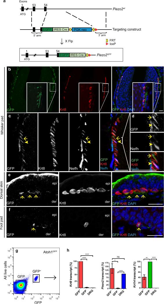
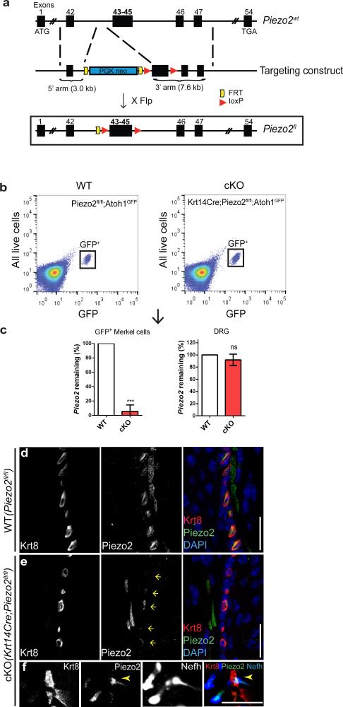
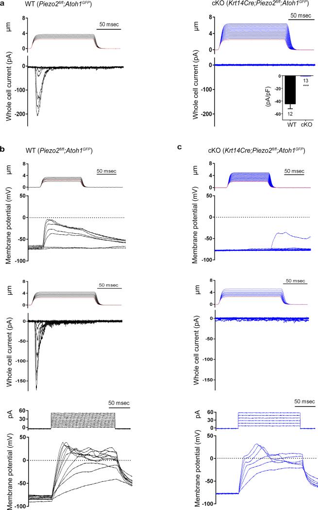
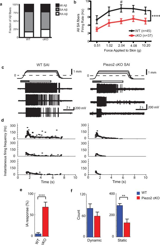
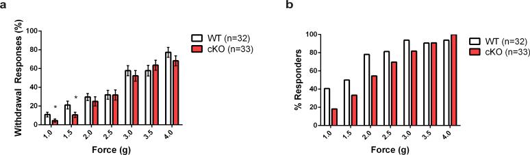
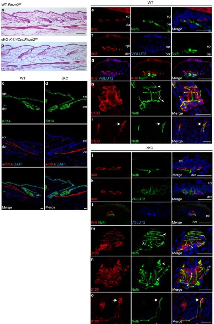
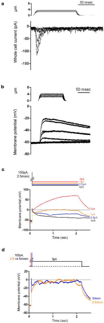
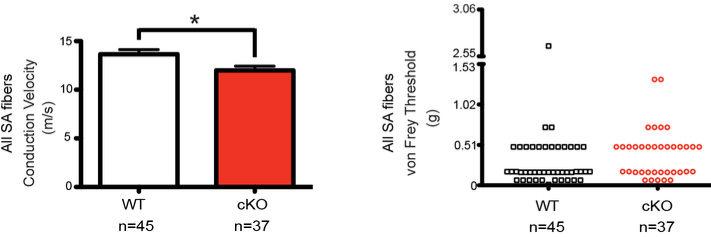
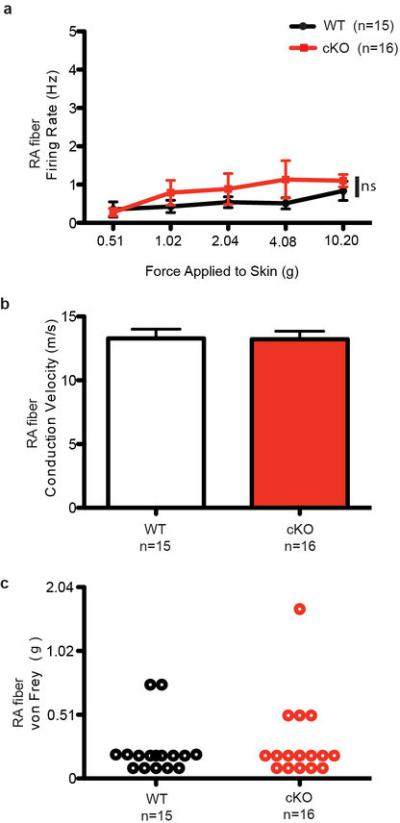
How we sense touch remains fundamentally unknown. The Merkel cell-neurite complex is a gentle touch receptor in the skin that mediates slowly adapting responses of Aβ sensory fibres to encode fine details of objects. This mechanoreceptor complex was recognized to have an essential role in sensing gentle touch nearly 50 years ago. However, whether Merkel cells or afferent fibres themselves sense mechanical force is still debated, and the molecular mechanism of mechanotransduction is unknown. Synapse-like junctions are observed between Merkel cells and associated afferents, and yet it is unclear whether Merkel cells are inherently mechanosensitive or whether they can rapidly transmit such information to the neighbouring nerve. Here we show that Merkel cells produce touch-sensitive currents in vitro. Piezo2, a mechanically activated cation channel, is expressed in Merkel cells. We engineered mice deficient in Piezo2 in the skin, but not in sensory neurons, and show that Merkel-cell mechanosensitivity completely depends on Piezo2. In these mice, slowly adapting responses in vivo mediated by the Merkel cell-neurite complex show reduced static firing rates, and moreover, the mice display moderately decreased behavioural responses to gentle touch. Our results indicate that Piezo2 is the Merkel-cell mechanotransduction channel and provide the first line of evidence that Piezo channels have a physiological role in mechanosensation in mammals. Furthermore, our data present evidence for a two-receptor-site model, in which both Merkel cells and innervating afferents act together as mechanosensors. The two-receptor system could provide this mechanoreceptor complex with a tuning mechanism to achieve highly sophisticated responses to a given mechanical stimulus.


Figures

Figure 1
Figure 1. Piezo2 expression in hairy and glabrous skin
a, A schematic diagram of the Piezo2GFP allele generation. Flp, flippase. b, GFP and Krt8 co-staining in the whisker follicle at a lower magnification. c, d, GFP, Krt8, and Nefh co-staining in the whisker follicle at a higher magnification. (d) shows a magnified view of the bracketed area in (c). Arrowheads mark the co-localization of GFP, Krt8, and Nefh. Note that in areas where Nefh+ fibers are missing, GFP and Krt8 still co-localize (arrows). e, f, GFP and Krt8 co-staining in a touch dome (e) and in glabrous skin (f). Arrows mark the position of Krt8+ Merkel cells. Scale bars b-f, 20 μm. epi, epidermis; der, dermis. g, h, A representative FACS plot (out of 12 experiments) of live epithelial cells isolated from Atoh1GFP skin (g) and qPCR analysis (n=4) of GFP+ and GFP cells and DRG isolated from Atoh1GFP mouse (h). Bars represent mean ± SEM. *P < 0.05; **P < 0.01; ****P < 0.0001; ns, not significantly different, unpaired t-test with Welch's correction.
Figure 2
Figure 2. Generation and characterization of skin-specific Piezo2 conditional knockout mice
a, A schematic diagram of the Piezo2fl allele generation. Flp, flippase. b, Representative FACS plots (out of 9 experiments) of live epithelial cells isolated from WT and cKO dorsal skin. c, qPCR (n=3) analysis showing Piezo2 levels in GFP+ cells and DRG of WT and cKO mice. Bars represent mean ± SEM. ***P < 0.001; ns, not significantly different, unpaired t-test with Welch's correction. d, e, Piezo2 and Krt8 co-staining in WT (d) and cKO (e) whisker pads. In (e), arrows mark the position of Krt8+ Merkel cells. f, Piezo2, Krt8, and Nefh co-staining in cKO whisker pad. Arrowheads mark the co-localization of Piezo2 and Nefh. Scar bars d-f, 20 μm.
Figure 3
Figure 3. Mechanically activated (MA) currents in Merkel cells depend on Piezo2
a, Representative traces of MA inward currents by a gentle poking stimulus in Merkel cells from WT (left) and cKO (right). The inset shows average MA whole-cell current density measured in WT and Piezo2 cKO Merkel cells. (CsCl intracellular) Bars represent mean ± SEM. ***P < 0.001, Student's t test. b, c, Representative traces (out of 6 experiments) from WT (b) and Piezo2 cKO (c) Merkel cells. Current clamp recordings displaying a change in membrane potential in response to gentle mechanical stimuli (top); subsequent displacement assays in voltage clamp mode (middle); current clamp recordings showing similar changes in membrane potential elicited by electrical stimulation (bottom). (K-gluconate intracellular) Red lines in stimulus traces indicate the displacement at which the probe visibly touched the cell.
Figure 4
Figure 4. Ex vivo skin-saphenous nerve recordings in WT and Piezo2 cKO mice
a, Proportions of mechanically insensitive (MI), slowly adapting (SA), and rapidly adapting (RA) Aβ fibers in WT (n=59) and Piezo2 cKO mice (n=66). The relatively low RA Aβ fiber % in both groups in comparison to previous studies is likely due to the genetic background of our animals. b, Firing rates of all SA Aβ fibers at increasing forces in WT and Piezo2 cKO. ****P < 0.0001; #P < 0.05, 2-way ANOVA with Bonferroni post-hoc analysis. c, Representative recordings from touch dome afferents in WT (left) vs Piezo2 cKO (right). Top traces show ramp-and-hold displacements at three magnitudes with corresponding spike trains below. Dashed line marks the point of skin contact (0 mm). Boxes indicate dynamic (dark gray: 1.5s after displacement command onset) and static phases for analysis (light gray: 4s after the beginning of hold command). d, Instantaneous firing frequencies of the responses in (c). e, Proportion of intermediately adapting (IA) responses to supra-threshold displacements in touch dome afferents. ****P <0.0001, Fisher's exact test. f, Maximum number of spikes in the dynamic and static response phases. **P ≤0.01, Student's t test. Bars represent mean ± SEM. For c-f, WT n=5, Piezo2 cKO n=6 afferents. Note that for a-b, recordings were made using a force-controlled mechanical stimulator; for c-f, a displacement control was used for the directed recordings of FM1-43-labeled afferents.
Figure 5
Figure 5. Gentle touch assay in WT and Piezo2 cKO mice
a, Percent paw withdrawal responses to gentle von Frey filament stimulation between 1.0 and 4.0 g forces in WT and Piezo2 cKO mice. Bars represent the mean ± SEM. *P < 0.05, Student's t test. b, Percent mice responding to von Frey filament stimulation in WT and Piezo2 cKO.
Extended Data Fig. 1
Extended Data Fig. 1. Validation of anti-Piezo2 antibody
a, Piezo2 detection by western blotting using anti-Piezo2 antibody (see full methods for antibody generation) in HEK293T cells overexpressing pIres2-EGFP (left lane), mPiezo1-pcDNA3.1(−)-Ires-EGFP (middle lane), and mPiezo2-sport6-Ires-EGFP (right lane). b, Piezo2 immunofluorescence in mPiezo2-sport6-Ires-EGFP-transfected HEK293T cells. The left panel shows EGFP epifluorescence in transfected cells, and the middle panel shows Piezo2 immunofluorescence in these same cells. c, d, Immunofluorescence of GFP and Piezo2 in adult Piezo2GFP reporter (c) and WT littermate (d) DRG. Piezo2 expression is observed in ~45.6 % of DRG neurons: 587 Piezo2+ expressers/1287 total neurons; 159 Piezo2high expressers/587 Piezo2+ expressers. Scale bars c, d, 100 μm.
Extended Data Fig. 2
Extended Data Fig. 2. GFP immunofluorescence in WT control andPiezo2GFP reporter mice
a, GFP, Krt8, and Nefh co-staining in WT littermate whisker follicle. b, c, GFP and Krt8 co-staining in WT littermate touch dome (b) and glabrous skin (c). Arrows mark the position of Krt8+ Merkel cells. d-h, GFP and Nefh co-staining in Piezo2GFP whisker follicle. (e-h) show magnified views of the bracketed area in (d). Arrows mark GFP expression only. Closed arrowheads mark the co-localization of GFP and Nefh. Scale bars a-h, 20 μm. epi, epidermis; der, dermis.
Extended Data Fig. 3
Extended Data Fig. 3. Generation of Piezo2-null allele (Piezo2) and characterization of Piezo2 constitutive knockout mice
a, A schematic diagram of Piezo2- allele generation. b, qPCR (n=2) showing Piezo2 levels in Piezo2wt/wt, Piezo2wt/−, and Piezo2−/− E19.5 lungs. Error bars represent mean ± SEM. **P < 0.01; ns, not significantly different, unpaired t-test with Welch's correction. c, d, Piezo2 immunofluorescence in WT littermate (c) and Piezo2−/− newborn DRG (d). Scale bars c, d, 100 μm.
Extended Data Fig. 4
Extended Data Fig. 4. Characterization of Piezo2fl/fl(WT) and Krt14Cre;Piezo2fl/fl(cKO) adult skin
a, b, H&E staining of WT and Piezo2 cKO dorsal skin. c, d, Immunofluorescence of Krt14 and a-SMA (alpha smooth muscle actin) in WT and cKO dorsal skin. e-g, j-l, Epidermal touch domes co-stained with Krt8, Nefh, and VGLUT2 (vesicular glutamate transporter 2, a marker for Merkel cells) in WT and cKO dorsal skin. h, m, n, Lanceolate endings and circumferential fibers co-stained with S100 (S100 calcium binding protein, a marker for Schwann cells) and Nefh in WT and cKO dorsal skin. Closed arrowheads mark circumferential fibers, and arrows mark lanceolate endings. i, o, Meissner's corpuscles co-stained with S100 and Nefh in WT and cKO footpads. Closed arrows mark Meissner's corpuscle. Scale bars, 100 μm (a, b), 20 μm (rest). epi, epidermis; der, dermis.
Extended Data Fig. 5
Extended Data Fig. 5. Current injections simulating Piezo2-induced currents produce prolonged depolarizations in Merkel cells in vitro
a, Representative traces of MA inward currents evoked by a gentle poking stimulus in a WT Merkel cell. A ramp (1 μm/msec)-and-hold displacement stimulus (0.25 μm increments) was applied to the cell in whole-cell voltage clamp configuration. The steady state current at the end of a 125 msec displacement was −6 ± 2pA (Vh= -80mV, n=15), 4% of the maximal current observed (−146 ± 29pA). b, Representative current clamp recordings from a WT Merkel cell, displaying a change in membrane potential in response to gentle mechanical stimuli. c, Piezo2-dependent currents were simulated by injecting short current pulses followed by different levels of long lasting but small current injections. In WT Merkel cells (n=4), membrane potential changes are elicited by applying a short (2.5 msec) 150 pA current injection followed by additional current injections of 0 pA (black), +0.5 pA (blue), +1 pA (orange) and +2 pA (red) from the bias holding current (-10 pA). In the absence of any continuous current, the membrane potential slowly decays after cessation of the initial 150 pA injection, consistent with a contribution of passive membrane properties (black trace). Importantly, long lasting depolarizations are observed when these short pulses are followed by very small current injections (0.5-1 pA, which are ~10-15 % of the average observed Piezo2 current remaining at the end of the 125 msec mechanical stimulation (Fig. 3b, see above)). d, In WT cells (n=4), membrane potential changes are elicited by a short 100 pA current injection followed by +3 pA (half the average Piezo2-dependent MA sustained current) from the bias holding current (−10 pA). Orange line, 2.5 msec initial pulse; Blue line, 5 msec initial pulse. These data indicate that long lasting Piezo2 channel activity at levels below that observed during mechanical stimulation (panel a and Fig. 3b) is crucial for sustained membrane depolarizations in Merkel cells. The high Rm of these cells can enable small current fluctuations to produce large voltage fluctuations.
Extended Data Fig. 6
Extended Data Fig. 6. Conduction velocity and von Frey thresholds of all SA Aβ afferents in WT and Piezo2 cKO mice
Conduction velocity (*P < 0.05, Student's t test) and von Frey thresholds (P = 0.0516, Mann-Whitney test) of all SA Aβ fibers from WT and Piezo2 cKO mice. Error bars represent mean ± SEM.
Extended Data Fig. 7
Extended Data Fig. 7. Characterization of RA Aβ afferents in WT and Piezo2 cKO mice
a, Firing rates of RA Aβ fibers in response to an increasing series of mechanical forces in WT and Piezo2 cKO mice. b, c, Conduction velocity (b) and von Frey thresholds (c) of RA Aβ fibers from WT and Piezo2 cKO mice.

Comment in

Similar articles

Cited by

References

    1. Abraira VE, Ginty DD. The sensory neurons of touch. Neuron. 2013;79:618–639. - PMC - PubMed
    1. Maksimovic S, Baba Y, Lumpkin EA. Neurotransmitters and synaptic components in the Merkel cell-neurite complex, a gentle-touch receptor. Annals of the New York Academy of Sciences. 2013;1279:13–21. - PMC - PubMed
    1. Chambers MR, Iggo A. Slowly-adapting cutaneous mechanoreceptors. The Journal of physiology. 1967;192:26–27. - PMC - PubMed
    1. Iggo A, Muir AR. The structure and function of a slowly adapting touch corpuscle in hairy skin. The Journal of physiology. 1969;200:763–796. - PMC - PubMed
    1. Johnson KO. The roles and functions of cutaneous mechanoreceptors. Current opinion in neurobiology. 2001;11:455–461. - PubMed

Publication types