. 2014 Jun;6(2):024111.
doi: 10.1088/1758-5082/6/2/024111. Epub 2014 Apr 10.

3D printing facilitated scaffold-free tissue unit fabrication

Affiliations

3D printing facilitated scaffold-free tissue unit fabrication

Yu Tan et al. Biofabrication. 2014 Jun.

Abstract

Tissue spheroids hold great potential in tissue engineering as building blocks to assemble into functional tissues. To date, agarose molds have been extensively used to facilitate fusion process of tissue spheroids. As a molding material, agarose typically requires low temperature plates for gelation and/or heated dispenser units. Here, we proposed and developed an alginate-based, direct 3D mold-printing technology: 3D printing microdroplets of alginate solution into biocompatible, bio-inert alginate hydrogel molds for the fabrication of scaffold-free tissue engineering constructs. Specifically, we developed a 3D printing technology to deposit microdroplets of alginate solution on calcium containing substrates in a layer-by-layer fashion to prepare ring-shaped 3D hydrogel molds. Tissue spheroids composed of 50% endothelial cells and 50% smooth muscle cells were robotically placed into the 3D printed alginate molds using a 3D printer, and were found to rapidly fuse into toroid-shaped tissue units. Histological and immunofluorescence analysis indicated that the cells secreted collagen type I playing a critical role in promoting cell-cell adhesion, tissue formation and maturation.


Figures

Figure 1
Figure 1
Schematic presentation of 3D alginate hydrogel printing on calcium-containing gelatin substrate. Adapted from the reference .
Figure 2
Figure 2
The size of alginate microdroplets (A=0.5, B=1.0, and C=1.5μl) printed on calcium containing gelatin substrates.
Figure 3
Figure 3
(a) Printing algorithm before optimization: printing microdroplets of alginate solution next to each other can lead to the coalescence of newly printed droplets. (b) Printing algorithm after optimization: printing microdroplets of alginate solution in 4 steps to to ensure that no printed droplet lands next to an un-gelled droplet and the final printed product can maintain a defined structure as designed.
Figure 4
Figure 4
Microdroplets facilitated 3D printing alginate hydrogels with different geometries (cube (a), square frame (b) and pyramid (c)).: the left is the schematic presentation of 3D printing algorithm used to print 3D structures shown in the right. Each layer was printed using an multi-step algorithm similar to the one shown in Figure 3. The optimal expected dimensions of 9.6 × 9.6 × 1.75 mm were based on a design of 9 mm × 9 mm in the X and Y dimension (measured from the dot center) and a Z dimension defined by 5 layers (d). The scale bar for (a),(b), and (c) is 1 mm. The scale bar for (d) is 2 mm. The blue, green, yellow, grey and red represent the 1st, 2nd, 3rd, 4th, and 5th layer of bioprinted alginate microdroplets, respectively.
Figure 5
Figure 5
Schematic presentation (a) and actual product (b) of 3D alginate hydrogel printing for tissue unit fabrication using vascular spheroids (i.e., containing smooth muscle cells and endothelial cells). Scale bar is 1mm
Figure 6
Figure 6
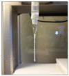
A picture showing the pasture pipette printing tip loaded with tissue spheroids for dispensing.
Figure 7
Figure 7
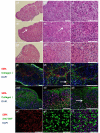
Histological and immunofluorescence analysis of the tissue units cultured for 4, 8 and 16 days. (a,b,c) H&E staining for tissue units cultured for 4 days at 10×, 20× and 40× magnification, respectively. (d,e,f) H&E staining for tissue units cultured for 8 days at 10×, 20× and 40× magnification, respectively. (g,h,i) H&E staining for tissue units cultured for 16 days at 10×, 20× and 40× magnification, respectively. (j,k,l) immunofluorescence analysis of tissue units cultured for 4 days at 20×, 40× and 63 × magnification. (m,n,o) immunofluorescence analysis of tissue units cultured for 16 days at 20×, 40× and 63 × magnification. (p,q,r) immunofluorescence analysis of tissue units cultured for 16 days at 40× magnification for smooth muscle actin, anti VWF and merge picture. Scale bar is 100μm

References

    1. Derby B. Printing and prototyping of tissues and scaffolds. Science. 2012;338:921–6. - PubMed
    1. Tasoglu S, Demirci U. Bioprinting for stem cell research. Trends in biotechnology. 2013;31:10–9. - PMC - PubMed
    1. Mironov V, Kasyanov V, Markwald RR. Organ printing: from bioprinter to organ biofabrication line. Current opinion in biotechnology. 2011;22:667–73. - PubMed
    1. Jakab K, Norotte C, Marga F, Murphy K, Vunjak-Novakovic G, Forgacs G. Tissue engineering by self-assembly and bio-printing of living cells. Biofabrication. 2010;2:022001. - PMC - PubMed
    1. Miller JS, Stevens KR, Yang MT, Baker BM, Nguyen DH, Cohen DM, et al. Rapid casting of patterned vascular networks for perfusable engineered three-dimensional tissues. Nature materials. 2012;11:768–74. - PMC - PubMed

Publication types