

Persistent salmonellosis causes pancreatitis in a murine model of infection
- PMID: 24717768
- PMCID: PMC3981665
- DOI: 10.1371/journal.pone.0092807
Persistent salmonellosis causes pancreatitis in a murine model of infection
Abstract
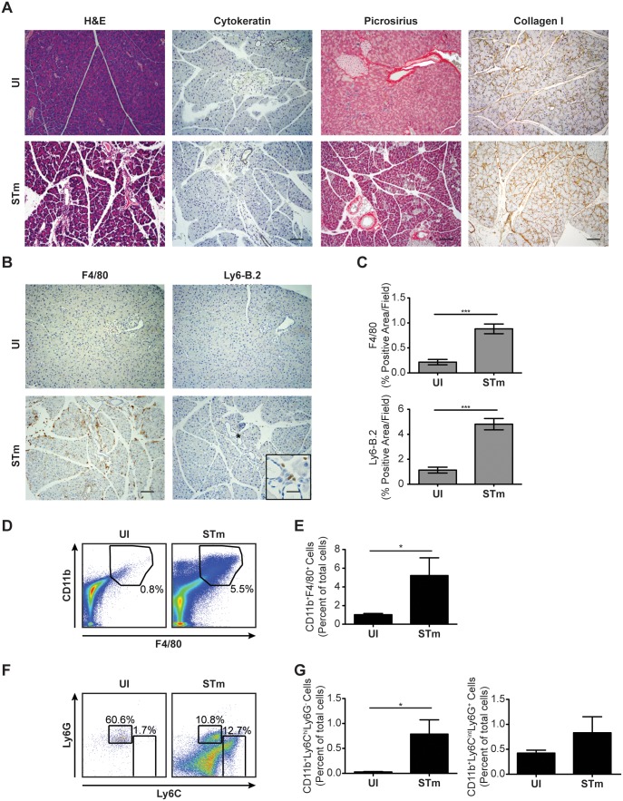
Pancreatitis, a known risk factor for the development of pancreatic ductal adenocarcinoma, is a serious, widespread medical condition usually caused by alcohol abuse or gallstone-mediated ductal obstruction. However, many cases of pancreatitis are of an unknown etiology. Pancreatitis has been linked to bacterial infection, but causality has yet to be established. Here, we found that persistent infection of mice with the bacterial pathogen Salmonella enterica serovar Typhimurium (S. Typhimurium) was sufficient to induce pancreatitis reminiscent of the human disease. Specifically, we found that pancreatitis induced by persistent S. Typhimurium infection was characterized by a loss of pancreatic acinar cells, acinar-to-ductal metaplasia, fibrosis and accumulation of inflammatory cells, including CD11b+ F4/80+, CD11b+ Ly6Cint Ly6G+ and CD11b+ Ly6Chi Ly6G- cells. Furthermore, we found that S. Typhimurium colonized and persisted in the pancreas, associated with pancreatic acinar cells in vivo, and could invade cultured pancreatic acinar cells in vitro. Thus, persistent infection of mice with S. Typhimurium may serve as a useful model for the study of pancreatitis as it relates to bacterial infection. Increased knowledge of how pathogenic bacteria can cause pancreatitis will provide a more integrated picture of the etiology of the disease and could lead to the development of new therapeutic approaches for treatment and prevention of pancreatitis and pancreatic ductal adenocarcinoma.
Conflict of interest statement
Figures


Similar articles
-
Numb regulates acinar cell dedifferentiation and survival during pancreatic damage and acinar-to-ductal metaplasia.Gastroenterology. 2013 Nov;145(5):1088-1097.e8. doi: 10.1053/j.gastro.2013.07.027. Epub 2013 Jul 25. Gastroenterology. 2013. PMID: 23891977 Free PMC article.
-
Pancreatic Acinar-to-Ductal Metaplasia and Pancreatic Cancer.Methods Mol Biol. 2019;1882:299-308. doi: 10.1007/978-1-4939-8879-2_26. Methods Mol Biol. 2019. PMID: 30378064
-
Inactivation of TGFβ receptor II signalling in pancreatic epithelial cells promotes acinar cell proliferation, acinar-to-ductal metaplasia and fibrosis during pancreatitis.J Pathol. 2016 Feb;238(3):434-45. doi: 10.1002/path.4666. Epub 2015 Nov 28. J Pathol. 2016. PMID: 26510396
-
Acinar-to-Ductal Metaplasia (ADM): On the Road to Pancreatic Intraepithelial Neoplasia (PanIN) and Pancreatic Cancer.Int J Mol Sci. 2023 Jun 9;24(12):9946. doi: 10.3390/ijms24129946. Int J Mol Sci. 2023. PMID: 37373094 Free PMC article. Review.
-
Acinar cells and the development of pancreatic fibrosis.Cytokine Growth Factor Rev. 2023 Jun-Aug;71-72:40-53. doi: 10.1016/j.cytogfr.2023.05.003. Epub 2023 May 26. Cytokine Growth Factor Rev. 2023. PMID: 37291030 Review.
Cited by
-
Inflammatory Monocytes Promote Granuloma-Mediated Control of Persistent Salmonella Infection.Infect Immun. 2022 Apr 21;90(4):e0007022. doi: 10.1128/iai.00070-22. Epub 2022 Mar 21. Infect Immun. 2022. PMID: 35311578 Free PMC article.
-
Limited Heme Oxygenase Contribution to Modulating the Severity of Salmonella enterica serovar Typhimurium Infection.Antioxidants (Basel). 2022 May 24;11(6):1040. doi: 10.3390/antiox11061040. Antioxidants (Basel). 2022. PMID: 35739937 Free PMC article.
-
The conspiracy of autophagy, stress and inflammation in acute pancreatitis.Curr Opin Gastroenterol. 2014 Sep;30(5):495-9. doi: 10.1097/MOG.0000000000000097. Curr Opin Gastroenterol. 2014. PMID: 25003605 Free PMC article. Review.
-
In trauma, expect the unexpected: a rare case of post-traumatic pancreatitis associated with salmonellosis and enterocolitis.BMJ Case Rep. 2018 Nov 8;2018:bcr2018226286. doi: 10.1136/bcr-2018-226286. BMJ Case Rep. 2018. PMID: 30413451 Free PMC article.
-
Mechanisms for the Invasion and Dissemination of Salmonella.Can J Infect Dis Med Microbiol. 2022 Jun 9;2022:2655801. doi: 10.1155/2022/2655801. eCollection 2022. Can J Infect Dis Med Microbiol. 2022. PMID: 35722038 Free PMC article. Review.
References
-
- Whitcomb DC (2004) Inflammation and Cancer V. Chronic pancreatitis and pancreatic cancer. Am J Physiol Gastrointest Liver Physiol 287: G315–319. - PubMed
-
- Fagenholz PJ, Castillo CF, Harris NS, Pelletier AJ, Camargo CA Jr (2007) Increasing United States hospital admissions for acute pancreatitis, 1988–2003. Ann Epidemiol 17: 491–497. - PubMed
-
- Tenner S, Sica G, Hughes M, Noordhoek E, Feng S, et al. (1997) Relationship of necrosis to organ failure in severe acute pancreatitis. Gastroenterology 113: 899–903. - PubMed
-
- Delrue LJ, De Waele JJ, Duyck PO (2010) Acute pancreatitis: radiologic scores in predicting severity and outcome. Abdom Imaging 35: 349–361. - PubMed
-
- Frey CF, Zhou H, Harvey DJ, White RH (2006) The incidence and case-fatality rates of acute biliary, alcoholic, and idiopathic pancreatitis in California, 1994–2001. Pancreas 33: 336–344. - PubMed
Publication types
MeSH terms
Substances
Grants and funding
LinkOut - more resources
Full Text Sources
Other Literature Sources
Medical
Research Materials
