Platypnoea-orthodeoxia syndrome: novel cause for a known condition
- PMID: 24717854
- PMCID: PMC3962980
- DOI: 10.1136/bcr-2013-201284
Platypnoea-orthodeoxia syndrome: novel cause for a known condition
Abstract
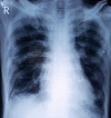
A 50-year-old man presented with dyspnoea while sitting, standing and walking but resolved completely in supine position. On cardiorespiratory examinations, fine crackles were noted over bibasal area. Chest X-ray showed bilateral reticulonodular shadows, restrictive pattern on spirometry, elevated alveolar arterial O2 gradient on arterial blood gas. High-resolution CT of the thorax revealed pattern as 'confident' or 'certain' radiographic diagnosis of idiopathic pulmonary fibrosis (IPF). Bubble-contrast echocardiography in recumbent, sitting and upright positions revealed no intracardiac (right to left shunt) or intrapulmonary shunts. This case highlights the necessity of awareness of this syndrome in cases of interstitial lung diseases (ILDs) also. Although 188 cases have been described thus far of platypnoea-orthodeoxia syndrome (P-OS) of various aetiologies, to the best of our knowledge, it is the first ever case of P-OS in ILD/IPF. Both lung bases were predominantly affected in this patient, platypnoea and orthodeoxia were attributed to areas of low/zero ventilation/perfusion (V/Q) ratio (zone 1 phenomena) as no other obvious explanation was found.
Figures


References
-
- Altman M, Robin ED. Platypnea (diffuse zone I phenomenon?). N Engl J Med 1969;281:1347–8 - PubMed
-
- Robin ED, Lamon D, Horn BR, et al. Platypnea related to orthodeoxia caused by true vascular lung shunts. N Engl J Med 1976;294:941–3 - PubMed
-
- Rodrigues P, Palma P, Sousa-Pereira L. Platypnea-orthodeoxia syndrome in review: defining a new disease? Cardiology 2012;123:15–23 - PubMed
-
- Burchell HB, Hemholz HF, Jr, Wood EH. Reflect orthostatic dyspnea associated with pulmonary hypotension. Am J Physiol 1949;159:563–4
-
- Seward JB, Hayes DL, Smith HC, et al. Platypnea-orthodeoxia: clinical profile, diagnostic workup, management and report of seven cases. Mayo Clin Proc 1984;59:221–31 - PubMed
Publication types
MeSH terms
Substances
LinkOut - more resources
Full Text Sources
Other Literature Sources
Medical
