

The stage-specific testicular germ cell apoptotic response to low-dose X-irradiation and 2,5-hexanedione combined exposure. I: Validation of the laser capture microdissection method for qRT-PCR array application
- PMID: 24717900
- PMCID: PMC4192118
- DOI: 10.1177/0192623314526319
The stage-specific testicular germ cell apoptotic response to low-dose X-irradiation and 2,5-hexanedione combined exposure. I: Validation of the laser capture microdissection method for qRT-PCR array application
Abstract
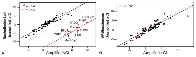
Over the past decade, laser capture microdissection (LCM) has grown as a tool for gene expression profiling of small numbers of cells from tumor samples and of specific cell populations in complex tissues. LCM can be used to study toxicant effects on selected cell populations within the testis at different stages of spermatogenesis. There are several LCM-related hurdles to overcome, including issues inherent to the method itself, as well as biases that result from amplifying the LCM-isolated RNA. Many technical issues associated with the LCM method are addressed here, including increasing RNA yield and obtaining more accurate quantification of RNA yields. We optimized the LCM method optimized to generate RNA quantities sufficient for quantitative reverse transcription polymerase chain reaction (qRT-PCR) array analysis without amplification and were able to validate the method through direct comparison of results from unamplified and amplified RNA from individual samples. The addition of an amplification step for gene expression studies using LCM RNA resulted in a bias, especially for low abundance transcripts. Although the amplification bias was consistent across samples, researchers should use caution when comparing results generated from amplified and unamplified LCM RNA. Here, we have validated the use of LCM-derived RNA with the qRT-PCR array, improving our ability to investigate cell-type and stage-specific responses to toxicant exposures.
Keywords: PCR array.; amplification; laser capture microdissection; testis.
© 2014 by The Author(s).
Conflict of interest statement
Competing Interests
Kim Boekelheide has funding from NIEHS, USEPA, and the American Chemistry Council. He is an occasional expert consultant for chemical and pharmaceutical companies, and owns stock in CytoSolv, an early stage biotechnology company developing a wound healing therapeutic.
Figures


Similar articles
-
The stage-specific testicular germ cell apoptotic response to low-dose radiation and 2,5-hexanedione combined exposure. II: qRT-PCR array analysis reveals dose dependent adaptive alterations in the apoptotic pathway.Toxicol Pathol. 2014 Dec;42(8):1229-37. doi: 10.1177/0192623314525689. Epub 2014 Mar 26. Toxicol Pathol. 2014. PMID: 24670816 Free PMC article.
-
Suppression of radiation-induced testicular germ cell apoptosis by 2,5-hexanedione pretreatment. III. Candidate gene analysis identifies a role for fas in the attenuation of X-ray-induced apoptosis.Toxicol Sci. 2010 Oct;117(2):466-74. doi: 10.1093/toxsci/kfq205. Epub 2010 Jul 8. Toxicol Sci. 2010. PMID: 20616204 Free PMC article.
-
Comparison of progestin transcriptional profiles in rat mammary gland using Laser Capture Microdissection and whole tissue-sampling.Exp Toxicol Pathol. 2013 Nov;65(7-8):949-60. doi: 10.1016/j.etp.2013.01.009. Epub 2013 Mar 7. Exp Toxicol Pathol. 2013. PMID: 23466250
-
Application of laser-capture microdissection to analysis of gene expression in the testis.Prog Histochem Cytochem. 2008;42(4):173-201. doi: 10.1016/j.proghi.2007.10.001. Prog Histochem Cytochem. 2008. PMID: 18243898 Review.
-
Laser-assisted microdissection in translational research: theory, technical considerations, and future applications.Appl Immunohistochem Mol Morphol. 2013 Jan;21(1):31-47. doi: 10.1097/PAI.0b013e31824d0519. Appl Immunohistochem Mol Morphol. 2013. PMID: 22495368 Review.
Cited by
-
Redox status of the testes and sperm of rats following exposure to 2,5-hexanedione.Redox Rep. 2016 Nov;21(6):239-47. doi: 10.1080/13510002.2015.1107312. Epub 2016 Feb 5. Redox Rep. 2016. PMID: 26818104 Free PMC article.
-
Is transcription in sperm stationary or dynamic?J Reprod Dev. 2017 Oct 18;63(5):439-443. doi: 10.1262/jrd.2016-093. Epub 2017 Aug 28. J Reprod Dev. 2017. PMID: 28845020 Free PMC article. Review.
References
-
- Campion SN, Houseman EA, Sandrof MA, Hensley JB, Sui Y, Gaido KW, Wu Z, Boekelheide K. Suppression of radiation-induced testicular germ cell apoptosis by 2,5-hexanedione pretreatment. II. Gene array analysis reveals adaptive changes in cell cycle and cell death pathways. Toxicological sciences: an official journal of the Society of Toxicology. 2010a;117:457–65. - PMC - PubMed
-
- Campion SN, Sandrof MA, Yamasaki H, Boekelheide K. Suppression of radiation-induced testicular germ cell apoptosis by 2,5-hexanedione pretreatment. III. Candidate gene analysis identifies a role for fas in the attenuation of X-ray-induced apoptosis. Toxicological sciences: an official journal of the Society of Toxicology. 2010b;117:466–74. - PMC - PubMed
-
- Caretti E, Devarajan K, Coudry R, Ross E, Clapper ML, Cooper HS, Bellacosa A. Comparison of RNA amplification methods and chip platforms for microarray analysis of samples processed by laser capture microdissection. Journal of cellular biochemistry. 2008;103:556–63. - PubMed
-
- Cheng L, Zhang S, MacLennan GT, Williamson SR, Davidson DD, Wang M, Jones TD, Lopez-Beltran A, Montironi R. Laser-assisted microdissection in translational research: theory, technical considerations, and future applications. Appl Immunohistochem Mol Morphol. 2013;21:31–47. - PubMed
Publication types
MeSH terms
Substances
Grants and funding
LinkOut - more resources
Full Text Sources
Other Literature Sources
Miscellaneous
