. 2014 Apr 9;9(4):e93987.
doi: 10.1371/journal.pone.0093987. eCollection 2014.

ESCRT-0 is not required for ectopic Notch activation and tumor suppression in Drosophila

Affiliations

ESCRT-0 is not required for ectopic Notch activation and tumor suppression in Drosophila

Emiliana Tognon et al. PLoS One. .

Abstract

Multivesicular endosome (MVE) sorting depends on proteins of the Endosomal Sorting Complex Required for Transport (ESCRT) family. These are organized in four complexes (ESCRT-0, -I, -II, -III) that act in a sequential fashion to deliver ubiquitylated cargoes into the internal luminal vesicles (ILVs) of the MVE. Drosophila genes encoding ESCRT-I, -II, -III components function in sorting signaling receptors, including Notch and the JAK/STAT signaling receptor Domeless. Loss of ESCRT-I, -II, -III in Drosophila epithelia causes altered signaling and cell polarity, suggesting that ESCRTs genes are tumor suppressors. However, the nature of the tumor suppressive function of ESCRTs, and whether tumor suppression is linked to receptor sorting is unclear. Unexpectedly, a null mutant in Hrs, encoding one of the components of the ESCRT-0 complex, which acts upstream of ESCRT-I, -II, -III in MVE sorting is dispensable for tumor suppression. Here, we report that two Drosophila epithelia lacking activity of Stam, the other known components of the ESCRT-0 complex, or of both Hrs and Stam, accumulate the signaling receptors Notch and Dome in endosomes. However, mutant tissue surprisingly maintains normal apico-basal polarity and proliferation control and does not display ectopic Notch signaling activation, unlike cells that lack ESCRT-I, -II, -III activity. Overall, our in vivo data confirm previous evidence indicating that the ESCRT-0 complex plays no crucial role in regulation of tumor suppression, and suggest re-evaluation of the relationship of signaling modulation in endosomes and tumorigenesis.


Conflict of interest statement

Competing Interests: The authors have declared that no competing interests exist.

Figures

Figure 1
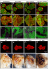
Figure 1. Hrs, Stam or Hrs, Stam double mutant tissue do not display altered tissue architecture.
(A–H) Epithelial morphology of mosaic FE cells (A–D) and eye discs (E–H) revealed by phalloidin staining to detect F-actin. Follicle cells of 5–7 stage egg chambers homozygous for the mutations (GFP-negative) show normal epithelial architecture compared to WT (GFP-positive). Eye disc cells homozygous for the mutations (GFP-negative) do not show any disruption of tissue architecture. (I–L) High magnification of a region of mosaic eye imaginal discs. Homozygous cells are marked by the absence of GFP. Apoptotic Caspase-3 (magenta) is activated cell autonomously in a subset of Hrs and Stam as well as Hrs, Stam mutant cells, compared to WT. (M–P) WT and predominantly mutant eye-antennal discs for the indicated gene stained with phalloidin revealed that Hrs, Stam mutant discs form morphologically normal eye-antennal discs. (Q–T) Adult eyes deriving from mosaic discs of the indicated genotype. Clones or WT (Q) or mutant cells (R–T) are marked by the absence of red pigment in bright field images indicating that mutant tissue can form photoreceptors.
Figure 2
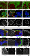
Figure 2. ESCRT-0 mutations lead to accumulation of ubiquitylated cargoes, as well as of Notch and Dome in endosomes.
(A–F) High magnification of a region of mosaic eye imaginal discs (A–D), or of FE (E–F) shows accumulation of ubiquitylated cargoes in mutant cells (GFP-negative), as revealed by an antibody against mono- and poly- ubiquitin chains (Ubi). High magnification of the boxed areas is shown in insets. (G–H) Mutant FE cells (GFP-negative) show accumulation of the Notch receptor. Notch receptor has been revealed using anti-NICD specific to the intracellular domain of Notch. Apical as well as intracellular accumulations of Notch ICD epitope is seen in Hrs and Stam FE mutant cells. High magnification of the boxed areas is shown in insets. (I–K) Co-localization with anti Notch ECD (NECD) or Notch ICD (NICD) and Avl, marking early endosomes, in mosaic eye imaginal discs. Notch ECD is mainly accumulated in early endosomes in GFP-negative mutant tissue. (L–L’) Mosaic eye imaginal discs were stained with Ubi and anti-Domeless (Dome). Hrs, Stam mutant cells (GFP-negative) accumulate ubiquitylated cargoes and moderate levels of Dome, compared to WT. (M–O) Endocytic trafficking assay with anti-Notch ECD to label Notch at the surface of living imaginal discs. In WT tissue, after labeling (0 hrs), Notch is present mostly at the apical surface of the cell. After a 5-hour chase (5 hrs) Notch is completely degraded in WT but still present in endosomes in Stam mutant discs, indicating that Notch is internalized but it is not degraded.
Figure 3
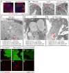
Figure 3. ESCRT-0 mutants do not affect endosomal maturation.
(A) Example of eye tissue almost completely homozygous for a WT (left) or mutant chromosome (right). WT cells are marked with GFP and represent 10% of the disc tissue. (B) Phase contrast images of cross-sections used for EM show the indicative regions used for ultrastructural analysis (pink boxes). Note the absence of monolayer architecture in sections of mutant tissues. (C–E) Electron micrograph of sections of eye disc tissue of the indicated genotype. A portion of the apical part of 2–3 epithelial cells above the level of the basal nuclei is shown. While MVEs (highlighted in red) are absent in Vps25 mutant cells, they are present in ESCRT-0 mutant cells. Quantification of MVE density, diameter, section area and ILV content is presented below each panel. (F–H) Incorporation of Lysotracker in mosaic discs. A single subapical confocal cross-section is shown in each panel, showing no difference in acidification in WT (GFP-positive) versus mutant cells. Labels are as follows: PM: peripodial membrane, DT: disc tissue, LU: Apical lumen, AJ: Adherens Junctions, ER: Endoplasmic Reticulum, GA: Golgi apparatus, MI: Mitocondrium, NU: Nucleus, GV: giant vacuoles, IS: interstitial space between unpolarized cells.
Figure 4
Figure 4. ESCRT-0 mutations do not alter Notch signaling in FE cells.
(A–F) Mosaic egg chambers at stages 5–7 of oogenesis stained to detect the Notch targets Hnt (A–C) and Cut (D–F) and f-Actin. Stam or Hrs Stam mutant cells are marked by the absence of GFP. In both Stam and Hrs Stam mutant FE cells, Hnt is normally expressed and Cut normally downregulated after stage 6, indicating no impairment of Notch signaling activation. (A’–F’) show single channels.

References

    1. Sigismund S, Confalonieri S, Ciliberto A, Polo S, Scita G, et al. (2012) Endocytosis and signaling: cell logistics shape the eukaryotic cell plan. Physiological reviews 92: 273–366. - PMC - PubMed
    1. Mellman I, Yarden Y (2013) Endocytosis and cancer. Cold Spring Harbor perspectives in biology 5: a016949. - PMC - PubMed
    1. Babst M, Katzmann D, Estepa-Sabal E, Meerloo T, Emr SD (2002) Escrt-III: an endosome-associated heterooligomeric protein complex required for mvb sorting. Developmental cell 3: 271–282. - PubMed
    1. Babst M, Katzmann D, Snyder W, Wendland B, Emr SD (2002) Endosome-associated complex, ESCRT-II, recruits transport machinery for protein sorting at the multivesicular body. Developmental cell 3: 283–289. - PubMed
    1. Katzmann D, Babst M, Emr SD (2001) Ubiquitin-dependent sorting into the multivesicular body pathway requires the function of a conserved endosomal protein sorting complex, ESCRT-I. Cell 106: 145–155. - PubMed

Publication types

MeSH terms

Substances