The clinical approach toward giant cell tumor of bone
- PMID: 24718514
- PMCID: PMC4012970
- DOI: 10.1634/theoncologist.2013-0432
The clinical approach toward giant cell tumor of bone
Abstract
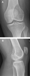
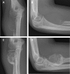
We provide an overview of imaging, histopathology, genetics, and multidisciplinary treatment of giant cell tumor of bone (GCTB), an intermediate, locally aggressive but rarely metastasizing tumor. Overexpression of receptor activator of nuclear factor κB ligand (RANKL) by mononuclear neoplastic stromal cells promotes recruitment of numerous reactive multinucleated giant cells. Conventional radiographs show a typical eccentric lytic lesion, mostly located in the meta-epiphyseal area of long bones. GCTB may also arise in the axial skeleton and very occasionally in the small bones of hands and feet. Magnetic resonance imaging is necessary to evaluate the extent of GCTB within bone and surrounding soft tissues to plan a surgical approach. Curettage with local adjuvants is the preferred treatment. Recurrence rates after curettage with phenol and polymethylmethacrylate (PMMA; 8%-27%) or cryosurgery and PMMA (0%-20%) are comparable. Resection is indicated when joint salvage is not feasible (e.g., intra-articular fracture with soft tissue component). Denosumab (RANKL inhibitor) blocks and bisphosphonates inhibit GCTB-derived osteoclast resorption. With bisphosphonates, stabilization of local and metastatic disease has been reported, although level of evidence was low. Denosumab has been studied to a larger extent and seems to be effective in facilitating intralesional surgery after therapy. Denosumab was recently registered for unresectable disease. Moderate-dose radiotherapy (40-55 Gy) is restricted to rare cases in which surgery would lead to unacceptable morbidity and RANKL inhibitors are contraindicated or unavailable.
Keywords: Curettage; Denosumab; GCTB; Giant cell tumor of bone; Histopathology; RANK ligand; Radiotherapy; Review.
Conflict of interest statement
Disclosures of potential conflicts of interest may be found at the end of this article.
Figures


Comment in
-
Giant cell tumor of bone.Oncologist. 2014 Nov;19(11):1207. doi: 10.1634/theoncologist.2014-0267. Oncologist. 2014. PMID: 25378541 Free PMC article.
-
In reply.Oncologist. 2014 Nov;19(11):1208. doi: 10.1634/theoncologist.2014-0332. Oncologist. 2014. PMID: 25378542 Free PMC article.
References
-
- Athanasou NA, Bansal M, Forsyth R, et al. Giant cell tumour of bone. In: Fletcher CD, Bridge JA, Hogendoorn PC, editors. WHO Classification of Tumours of Soft Tissue and Bone. Lyon, France: IARC Press; 2013. pp. 321–324.
-
- Balke M, Schremper L, Gebert C, et al. Giant cell tumor of bone: Treatment and outcome of 214 cases. J Cancer Res Clin Oncol. 2008;134:969–978. - PubMed
-
- Campanacci M, Baldini N, Boriani S, et al. Giant-cell tumor of bone. J Bone Joint Surg Am. 1987;69:106–114. - PubMed
-
- Forsyth R, Jundt G. Giant cell lesion of the small bones. In: Fletcher CD, Bridge JA, Hogendoorn PC, editors. WHO Classification of Tumours of Soft Tissue and Bone. Lyon, France: IARC Press; 2013. p. 320.
-
- Siebenrock KA, Unni KK, Rock MG. Giant-cell tumour of bone metastasising to the lungs. A long-term follow-up. J Bone Joint Surg Br. 1998;80:43–47. - PubMed
Publication types
MeSH terms
Substances
LinkOut - more resources
Full Text Sources
Other Literature Sources
Medical

