. 2014 Apr 9;9(4):e94380.
doi: 10.1371/journal.pone.0094380. eCollection 2014.

Nesprins: tissue-specific expression of epsilon and other short isoforms

Affiliations

Nesprins: tissue-specific expression of epsilon and other short isoforms

Nguyen Thuy Duong et al. PLoS One. .

Abstract

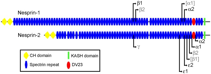
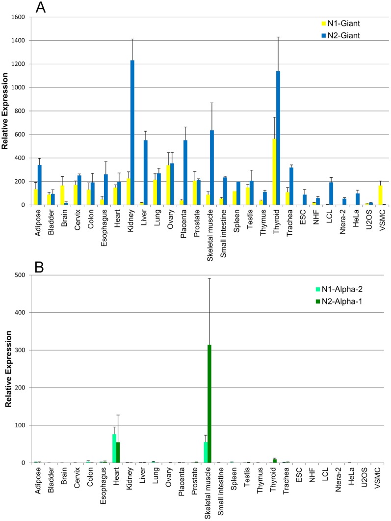
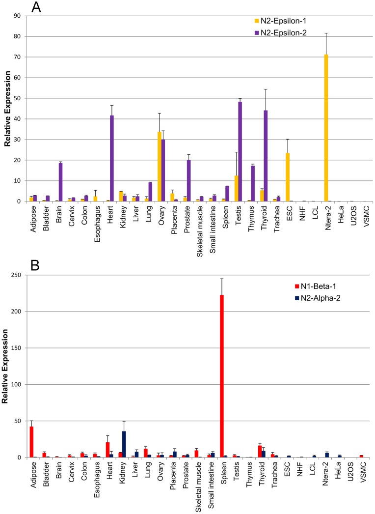
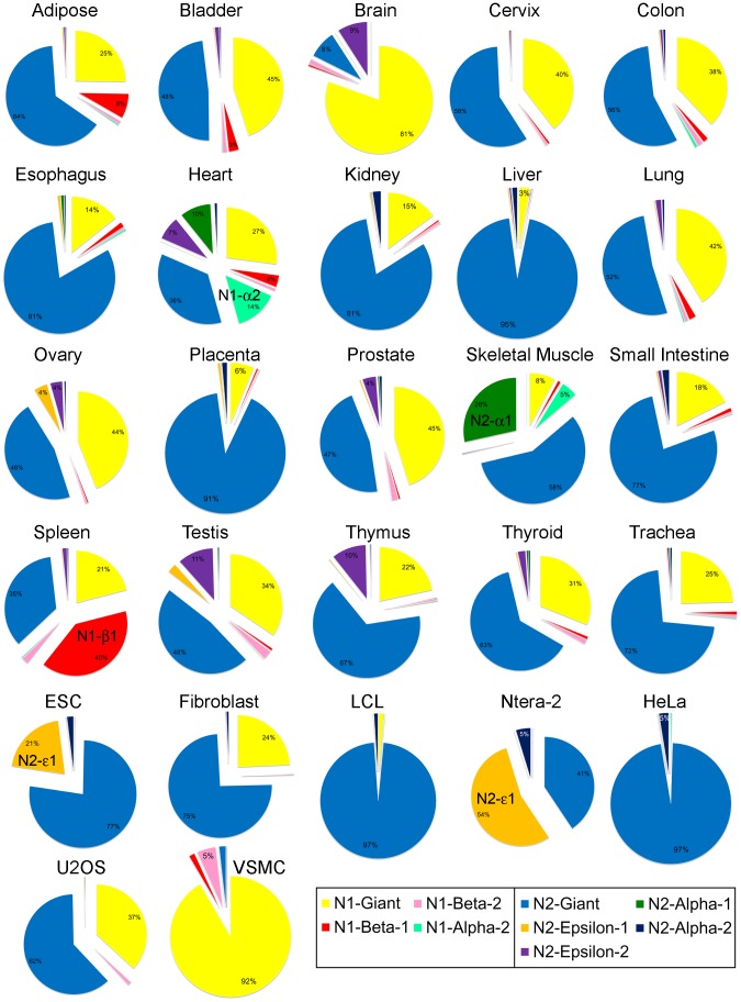
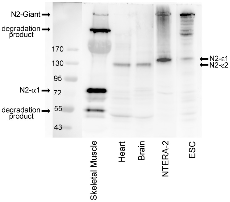
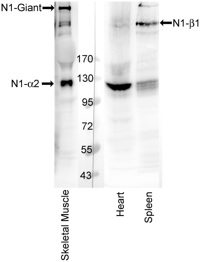
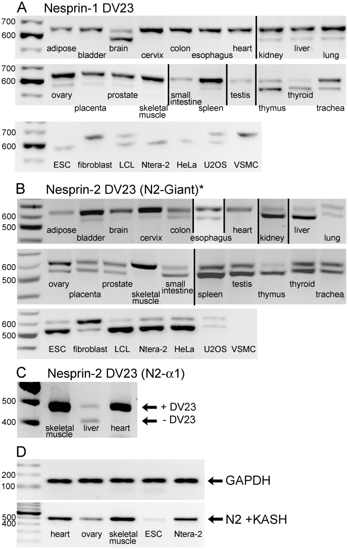
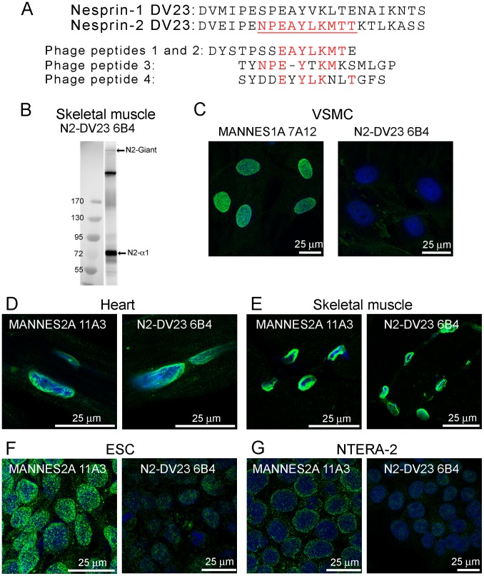
Nesprin-1-giant and nesprin-2-giant regulate nuclear positioning by the interaction of their C-terminal KASH domains with nuclear membrane SUN proteins and their N-terminal calponin-homology domains with cytoskeletal actin. A number of short isoforms lacking the actin-binding domains are produced by internal promotion. We have evaluated the significance of these shorter isoforms using quantitative RT-PCR and western blotting with site-specific monoclonal antibodies. Within a complete map of nesprin isoforms, we describe two novel nesprin-2 epsilon isoforms for the first time. Epsilon isoforms are similar in size and structure to nesprin-1-alpha. Expression of nesprin isoforms was highly tissue-dependent. Nesprin-2-epsilon-1 was found in early embryonic cells, while nesprin-2-epsilon-2 was present in heart and other adult tissues, but not skeletal muscle. Some cell lines lack shorter isoforms and express only one of the two nesprin genes, suggesting that either of the giant nesprins is sufficient for basic cell functions. For the first time, localisation of endogenous nesprin away from the nuclear membrane was shown in cells where removal of the KASH domain by alternative splicing occurs. By distinguishing between degradation products and true isoforms on western blots, it was found that previously-described beta and gamma isoforms are expressed either at only low levels or with a limited tissue distribution. Two of the shortest alpha isoforms, nesprin-1-alpha-2 and nesprin-2-alpha-1, were found almost exclusively in cardiac and skeletal muscle and a highly conserved and alternatively-spliced exon, available in both nesprin genes, was always included in these tissues. These "muscle-specific" isoforms are thought to form a complex with emerin and lamin A/C at the inner nuclear membrane and mutations in all three proteins cause Emery-Dreifuss muscular dystrophy and/or inherited dilated cardiomyopathy, disorders in which only skeletal muscle and/or heart are affected.


Conflict of interest statement

Competing Interests: The authors have declared that no competing interests exist.

Figures

Figure 1
Figure 1. Short forms of nesprin-1 and nesprin-2.
Pictorial representation of nesprin-1-giant and nesprin-2-giant with the N-terminal start points of the smaller isoforms indicated by black lines and arrows. Isoforms that we found present at low levels are labelled in grey. Those isoforms that were barely detectable, or undetectable, are labelled in grey with parenthesis. (See Results).
Figure 2
Figure 2. Tissue-specific expression of giant and alpha isoforms of nesprins.
Quantitative PCR to show mRNA expression of nesprin isoforms relative to the expression of two endogenous house-keeping controls. Charts represent the mean relative expression ± SEM, measured in cDNA preparations from 20 human tissues and 7 cell lines. These values are given in Tables S3 and S4 in File S1. Bar charts show relative expression of (A) nesprin-1-giant and nesprin-2-giant, and (B) nesprin-1-alpha-2 and nesprin-2-alpha-1.
Figure 3
Figure 3. Tissue-specific expression of nesprin-1-beta-1, nesprin-2-alpha-2 and the two nesprin-2-epsilon isoforms.
Quantitative PCR to show mRNA expression of nesprin isoforms relative to the expression of two endogenous house-keeping controls. Charts represent the mean relative expression ± SEM, measured in cDNA preparations from 20 human tissues and 7 cell lines. These values are given in Tables S3 and S4 in File S1. Bar charts show relative expression of (A) nesprin-2-epsilon-1 and nesprin-2-epsilon-2, and (B) nesprin-1-beta-1 and nesprin-2-alpha-2.
Figure 4
Figure 4. Relative abundance of nesprin isoform mRNAs in human tissues and cell lines.
Exploded pie charts show the relative abundance of nesprin isoform mRNA, relative to total nesprin-1 and nesprin-2 mRNA in cDNA preparations from 20 human tissues and 7 human cell lines.
Figure 5
Figure 5. Evidence at the protein level for nesprin-2-epsilon-1 in Ntera-2 and ESC cells and for nesprin-2-epsilon-2 in heart and brain.
Western blot for nesprin-2 in human tissues and cell lines using antibody against the C-terminal region of nesprin-2 (MANNES2A 11A3). Bands of approximate size of nesprin-2-epsilon-1 (122 kD) were detected in Ntera-2 and ESC and bands the size of nesprin-2-epsilon-2 (103 kD) were detected in brain and heart. Nesprin-2-alpha-1 was observed in skeletal muscle, but epsilon isoforms were not detected. Absence of mRNA (see Table S4 in File S1), indicates that putative nesprin-2-gamma (377 kD) and nesprin-2-alpha-2 (47 kD) bands on the skeletal muscle western blot, are likely to be degradation products.
Figure 6
Figure 6. Evidence at the protein level for nesprin-1-beta-1 in spleen and for nesprin-1-alpha-2 in cardiac and skeletal muscle.
Western blot for nesprin-1 in tissues using antibody against the C-terminal region of nesprin-1 (MANNES1E 8C3). A band the size of nesprin-1-beta-1 (383 kD) was detected in spleen and bands of nesprin-1-alpha-2 (111 kD) were detected in skeletal muscle and heart.
Figure 7
Figure 7. Tissue-dependent alternative splicing of the highly conserved DV23 exon of nesprin-1 and nesprin-2 and the KASH domain of nesprin-2.
Products of conventional PCR were separated on agarose gels. The 27 human cDNA samples, as shown in Table 1, were used in A and B. For DV23, the upper band contained the DV23 region and the lower band lacked the DV23 region. Specific primer pairs spanned: (A) The nesprin-1 DV23 region of nesprin-1-giant and other nesprin-1 isoforms, (B) the nesprin-2 DV23 region of nesprin-2-giant (which included other nesprin-2 isoforms except nesprin-2-alpha-1) and (C) the nesprin-2 DV23 region of only the nesprin-2-alpha-1 isoform. Vertical black lines indicate images from different gels that have been compiled. (D) PCR was used to detect the presence of nesprin-2 KASH, along with GAPDH control in 5 cDNA samples.
Figure 8
Figure 8. Immunolocalisation of isoforms lacking the DV23 region and/or the KASH domain.
(A) Alignment of the 23 amino acid sequences of the DV23 exon of nesprin-1 and nesprin-2. The nesprin-2 DV23 peptide was the immunogen for mAb production. The epitope of the mAbs was mapped to the sequence shown in red and underlined. Sequences of the four 15-mer peptides pulled out of the phage-display peptide library are shown aligned below the nesprin-2 DV23 sequence, with matching amino acids shown in red. (B) The mAb N2-DV23 6B4 recognised bands of nesprin-2-giant and nesprin-2-alpha-1 in western blot of skeletal muscle (the intermediate band is a likely degradation product of nesprin-2-giant). (C) The 6B4 mAb against nesprin-2 DV23 does not cross-react with nesprin-1 DV23. VSMC (which contain nesprin-1 with DV23 but no nesprin-2) showed nuclear staining for nesprin-1 with MANNES1A but not with 6B4. Immunofluorescent staining with mAbs MANNES2A and N2-DV23 6B4 was equally strong on both cardiac muscle nuclear rim (D) and skeletal muscle nuclear rim (E). However, MANNES2A was much stronger than N2-DV23 6B4 on both ESC (F) and Ntera-2 cells (G), since the DV23 exon is mainly excluded in these cells (see Fig. 7B). Furthermore, the MANNES1A mAb is largely nucleoplasmic in ESC, which may be because the nesprin-2 KASH domain is also largely excluded in this cell type (see Fig. 7D).

Similar articles

Cited by

References

    1. Apel ED, Lewis RM, Grady RM, Sanes JR (2000) Syne-1, a dystrophin- and klarsicht-related protein associated with synaptic nuclei at the neuromuscular junction. J Biol Chem 275: 31986–31995. - PubMed
    1. Zhang Q, Skepper JN, Yang F, Davies JD, Hegyi L, et al. (2001) Nesprins: a novel family of spectrin-repeat-containing proteins that localize to the nuclear membrane in multiple tissues. J Cell Sci 114: 4485–98. - PubMed
    1. Zhen YY, Libotte T, Munck M, Noegel AA, Korenbaum E (2002) NUANCE, a giant protein connecting the nucleus and actin cytoskeleton. J Cell Sci 115: 3207–3222. - PubMed
    1. Zhang Q, Ragnauth CD, Skepper JN, Worth NF, Warren DT, et al. (2005) Nesprin-2 is a multi-isomeric protein that binds lamin and emerin at the nuclear envelope and forms a subcellular network in skeletal muscle. J Cell Sci 118: 673–687. - PubMed
    1. Mislow JM, Kim MS, Davis DB, McNally EM (2002) Myne-1, a spectrin repeat transmembrane protein of the myocyte inner nuclear membrane, interacts with lamin A/C. J Cell Sci 115: 61–70. - PubMed

Publication types

MeSH terms

LinkOut - more resources