. 2014 Apr 9;34(15):5083-8.
doi: 10.1523/JNEUROSCI.4662-13.2014.

Differential stability of PNS and CNS nodal complexes when neuronal neurofascin is lost

Affiliations

Differential stability of PNS and CNS nodal complexes when neuronal neurofascin is lost

Anne Desmazieres et al. J Neurosci. .

Abstract

Fast, saltatory conduction in myelinated nerves requires the clustering of voltage-gated sodium channels (Nav) at nodes of Ranvier in a nodal complex. The Neurofascin (Nfasc) gene encodes neuronal Neurofascin 186 (Nfasc186) at the node and glial Neurofascin 155 at the paranode, and these proteins play a key role in node assembly. However, their role in the maintenance and stability of the node is less well understood. Here we show that by inducible ablation of Nfasc in neurons in adult mice, Nfasc186 expression is reduced by >99% and 94% at PNS and CNS nodes, respectively. Gliomedin and NrCAM at PNS and brevican at CNS nodes are largely lost with neuronal neurofascin; however, Nav at nodes of Ranvier persist, albeit with ∼40% reduction in expression levels. βIV Spectrin, ankyrin G, and, to a lesser extent, the β1 subunit of the sodium channel, are less affected at the PNS node than in the CNS. Nevertheless, there is a 38% reduction in PNS conduction velocity. Loss of Nfasc186 provokes CNS paranodal disorganization, but this does not contribute to loss of Nav. These results show that Nav at PNS nodes are still maintained in a nodal complex when neuronal neurofascin is depleted, whereas the retention of nodal Nav in the CNS, despite more extensive dissolution of the complex, suggests a supportive role for the partially disrupted paranodal axoglial junction in selectively maintaining Nav at the CNS node.

Keywords: myelination; neurofascin; nodes of Ranvier; sodium channels.


Figures

Figure 1.
Figure 1.
Characterization of Nfasc186 deletion and preservation of myelin in the TCE/Nfascfl/− model. Control (Nfascfl/−) and mutant (TCE/Nfascfl/−) mice received intraperitoneal injections of tamoxifen to induce the recombination of the Nfasc floxed allele. A, Western blotting of peripheral nerve and spinal cord lysates from control and mutant mice 16 weeks post-tamoxifen shows substantial loss of Nfasc186, while glial Nfasc155 is unaffected. Tubulin was the loading control. B, Immunofluorescence of teased quadriceps nerves (PNS) and ventral funiculi of cervical spinal cord (CNS) from Nfascfl/− and TCE/Nfascfl/− mice shows the disappearance of nodal Nfasc186 at 8 weeks (wk) and 16 weeks post-tamoxifen in both PNS and CNS. The Pan Nfasc antibody recognizes both Nfasc186 and Nfasc155. Scale bar, 3 μm. C, Quantitation of nodal area immunoreactive for Nfasc186 in mutant quadriceps nerves and ventral funiculi at 8 weeks and 16 weeks post-tamoxifen as a percentage of control. D, Toluidine blue-stained semithin transverse sections of quadriceps nerves (PNS) and ventral funiculi of cervical spinal cord (CNS) from Nfascfl/− and TCE/Nfascfl/− mice 16 weeks post-tamoxifen show no obvious alteration of myelin in myelinated axons. Scale bar, 10 μm. E, Western blotting of peripheral nerve and spinal cord lysates 16 weeks post-tamoxifen shows normal amounts of the myelin basic proteins (MBP) in Nfascfl/− and TCE/Nfascfl/− mice. Actin was the loading control.
Figure 2.
Figure 2.
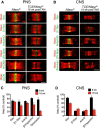
Loss of Nfasc186 leads to differential loss of nodal proteins in the PNS and CNS. A, B, Immunofluorescence of quadriceps nerves (PNS; A) and spinal cord ventral funiculi (CNS; B) from control (Nfascfl/−) and mutant (TCE/Nfascfl/−) mice at 16 weeks (wk) post-tamoxifen shows disruption of nodal components following inducible deletion of Nfasc186. βIV Spec, βIV Spectrin; GLDN, gliomedin. C, D, The quantitation of nodal disruption is shown for PNS (C) and CNS (D) where mean nodal areas ± SEM for the mutant are expressed as percentage of control mean nodal area at 8 and 16 weeks post-tamoxifen treatment. Scale bar, 3 μm.
Figure 3.
Figure 3.
CNS paranodal and juxtaparanodal proteins are mislocalized following Nfasc186 deletion. A, Immunofluorescence of teased ventral funiculi from Nfascfl/− and TCE/Nfascfl/− mice 16 weeks (wk) post-tamoxifen treatment shows disrupted Caspr staining and relocalization of voltage-gated potassium channel (Kv) at the paranodes of mutants. Scale bar, 3 μm. B, Western blotting of spinal cord lysates from control and mutant mice 16 weeks post-tamoxifen shows no reduction in Caspr or contactin in mutant nerves. Actin was the loading control. C, The percentage of paranodes displaying invasive Kv immunoreactivity was measured in wild-type (Nfasc+/+), control (Nfascfl/−), and mutant (TCE/Nfascfl/−) mice 16 weeks post-tamoxifen (wild-type mice were the same age but not treated with tamoxifen) and are displayed as means ± SEM. There is a significant increase in disrupted paranodes in mutants compared with controls (one-way ANOVA followed by Tukey's multiple-comparison test; **p = 0.0001; n = 3, 120 paranodes per condition). ns, not significant. D, The mean areas of Nav immunoreactivity were measured when either paranodes were intact (Kv restricted to juxtaparanodes) or disrupted (Kv invasion of paranodes). In the control and the mutant, there is no significant difference (ns) in Nav localization at the nodes when paranodes are either intact or disrupted (mean ± SEM, one-way ANOVA followed by Tukey's multiple-comparison tests; p = 0.01, n = 3, minimum 90 nodes).
Figure 4.
Figure 4.
Diagram contrasting the sequence of events when Nfasc186 is lost in the PNS and CNS. The intensity of the colors indicates when proteins are diminished but not completely lost.

Similar articles

Cited by

References

    1. Amor V, Feinberg K, Eshed-Eisenbach Y, Vainshtein A, Frechter S, Grumet M, Rosenbluth J, Peles E. Long-term maintenance of Na+ channels at nodes of Ranvier depends on glial contact mediated by gliomedin and NrCAM. J Neurosci. 2014;34:5089–5098. - PMC - PubMed
    1. Bhat MA, Rios JC, Lu Y, Garcia-Fresco GP, Ching W, St Martin M, Li J, Einheber S, Chesler M, Rosenbluth J, Salzer JL, Bellen HJ. Axon-glia interactions and the domain organization of myelinated axons requires neurexin IV/Caspr/Paranodin. Neuron. 2001;30:369–383. doi: 10.1016/S0896-6273(01)00294-X. - DOI - PubMed
    1. Boyle ME, Berglund EO, Murai KK, Weber L, Peles E, Ranscht B. Contactin orchestrates assembly of the septate-like junctions at the paranode in myelinated peripheral nerve. Neuron. 2001;30:385–397. doi: 10.1016/S0896-6273(01)00296-3. - DOI - PubMed
    1. Charles P, Tait S, Faivre-Sarrailh C, Barbin G, Gunn-Moore F, Denisenko-Nehrbass N, Guennoc AM, Girault JA, Brophy PJ, Lubetzki C. Neurofascin is a glial receptor for the paranodin/Caspr-contactin axonal complex at the axoglial junction. Curr Biol. 2002;12:217–220. doi: 10.1016/S0960-9822(01)00680-7. - DOI - PubMed
    1. Chen C, Westenbroek RE, Xu X, Edwards CA, Sorenson DR, Chen Y, McEwen DP, O'Malley HA, Bharucha V, Meadows LS, Knudsen GA, Vilaythong A, Noebels JL, Saunders TL, Scheuer T, Shrager P, Catterall WA, Isom LL. Mice lacking sodium channel beta1 subunits display defects in neuronal excitability, sodium channel expression, and nodal architecture. J Neurosci. 2004;24:4030–4042. doi: 10.1523/JNEUROSCI.4139-03.2004. - DOI - PMC - PubMed

Publication types

LinkOut - more resources