. 2014 Apr 9;34(15):5089-98.
doi: 10.1523/JNEUROSCI.4752-13.2014.

Long-term maintenance of Na+ channels at nodes of Ranvier depends on glial contact mediated by gliomedin and NrCAM

Affiliations

Long-term maintenance of Na+ channels at nodes of Ranvier depends on glial contact mediated by gliomedin and NrCAM

Veronique Amor et al. J Neurosci. .

Abstract

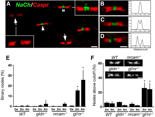
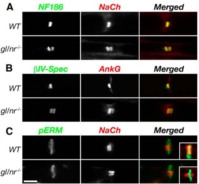
Clustering of Na(+) channels at the nodes of Ranvier is coordinated by myelinating glia. In the peripheral nervous system, axoglial contact at the nodes is mediated by the binding of gliomedin and glial NrCAM to axonal neurofascin 186 (NF186). This interaction is crucial for the initial clustering of Na(+) channels at heminodes. As a result, it is not clear whether continued axon-glial contact at nodes of Ranvier is required to maintain these channels at the nodal axolemma. Here, we report that, in contrast to mice that lack either gliomedin or NrCAM, absence of both molecules (and hence the glial clustering signal) resulted in a gradual loss of Na(+) channels and other axonal components from the nodes, the formation of binary nodes, and dysregulation of nodal gap length. Therefore, these mice exhibit neurological abnormalities and slower nerve conduction. Disintegration of the nodes occurred in an orderly manner, starting with the disappearance of neurofascin 186, followed by the loss of Na(+) channels and ankyrin G, and then βIV spectrin, a sequence that reflects the assembly of nodes during development. Finally, the absence of gliomedin and NrCAM led to the invasion of the outermost layer of the Schwann cell membrane beyond the nodal area and the formation of paranodal-like junctions at the nodal gap. Our results reveal that axon-glial contact mediated by gliomedin, NrCAM, and NF186 not only plays a role in Na(+) channel clustering during development, but also contributes to the long-term maintenance of Na(+) channels at nodes of Ranvier.

Keywords: NrCAM; Schwann; gliomedin; myelin; neurofascin; node of Ranvier.


Figures

Figure 1.
Figure 1.
Absence of gliomedin and NrCAM results in motor abnormalities. A, RT-PCR analysis of sciatic nerve mRNA using primer pairs specific to gliomedin and NrCAM revealed the absence of both transcripts in double homozygous mutant mice (gl/nr−/−). Primers for actin were used as controls. Size markers are given in base pairs. B, Gliomedin and NrCAM are undetectable in sciatic nerves of the double mutant mice. Teased sciatic nerves isolated from P60 mice of the indicated genotypes were immunolabeled using antibodies to gliomedin (Gldn), NrCAM, and P0 protein (P0). The location of the node in the double mutant is marked with an asterisk. Scale bar, 10 μm. C, Double mutant mice exhibit abnormal clasping of their hind limbs when suspended by their tails. Note the normal hind legs spreading of the single mutant animals. D, Adult double mutant mice (nrcam−/−/gldn−/−) exhibit a shorter latency to fall when left to hang from a horizontal rod by their forelimbs. Note that >80% of the double mutants fell after <5 s compared with wild-type mice, which stayed on the bar for more than a minute (n = 8). E, The double mutants stumble when placed on horizontal bars, whereas their single mutant littermates were undistinguishable from wild-type mice (nrcam−/−, bottom, and data not shown). F, Compound action potentials recorded from P15 mice show a reduction in nerve conduction velocity in the double but not single mutant mice. Error bars indicate SEM of n > 7 mice per genotype; p < 0.001. G, Electron microscopy images of sciatic nerve cross sections obtained from P120 wild-type (WT) mice, NrCAM-null (nrcam−/−), gliomedin-null (gldn−/−), or double mutant (gldn−/−/nrcam−/−) mice showing normal myelin. Scale bar, 10 μm.
Figure 2.
Figure 2.
Disappearance of nodal Na+ channels in double gldn−/−/nrcam−/− mutant sciatic nerves. A, Immunolabeling of teased sciatic nerve fibers isolated from adult (P120) double mutant, showing nodes of Ranvier that lack Na+ channels (asterisks). Immunolabeling using antibodies to Caspr and neurofilament (NF-H) was performed to label the paranodal junction and the axon, respectively. B, Quantitative analysis of sciatic nerves isolated at P15, P60, and P120 from wild-type (WT), gliomedin-null (gldn−/−), NrCAM-null (nrcam−/−), and double mutant (gldn−/−/nrcam−/−) mice. Percent of nodes lacking Na+ channels is shown in gray; nodes containing Na+ channels are in black. More than 300 nodes from 3–5 animals per genotype were analyzed, *p < 0.0001. Scale bar, 10 μm.
Figure 3.
Figure 3.
Double mutant mice exhibit binary Na+ channel clusters and widening of the nodal gap. A, Immunofluorescence labeling of teased sciatic nerve isolated from a P120 double mutant mouse using antibodies to Na+ channels (NaCh) and Caspr. At this age, Na+ channels were either present (arrowhead), absent (arrows), or appeared as binary clusters (two small arrowheads) at the nodal gap. BD, Additional examples of binary Na+ channel clusters in the double mutant (Na+ channels, green; Caspr, red). A line scan representing the intensity of the Na+ channel fluorescence signal (arbitrary units) along the nodal area is shown on the right of each panel. E, Increased appearance of binary nodal Na+ channels with age. Quantitation of the number of nodes containing binary Na+ channel clusters in sciatic nerves of wild-type (WT), single (gldn−/−, or nrcam−/−), or double (gl/nr−/−) mutant mice at P15 (2w), P60 (2m), and P120 (4m). Data are presented as a percentage of all Na+-channel-positive nodes. Error bars indicate SD of n = 3–5 animals per age per genotype (300 nodes per group), *p < 0.001. F, Double mutant nodes of Ranvier are longer. P60 or P120 sciatic nerves, isolated from the indicated genotypes, were labeled using antibodies to Caspr to demarcate the nodal gap. An asterisk marks a longer nodal gap in the double mutant. The percent of nodes that are longer than 1.3 μm and 1.6 μm in P60 and P120 mice, respectively, is shown. Error bars indicate SD of n = 3–5 animals per genotype per age (counted at least 300 sites per group), *p < 0.005. Scale bars, 5 μm.
Figure 4.
Figure 4.
Molecular composition of the nodes in the absence of gliomedin and NrCAM. Immunolabeling of teased sciatic nerve fibers isolated from adult wild-type (WT) and double mutant mice (gl−/−/nr−/−) using antibodies to neurofascin 186 (NF186) and Na+ channels (NaCh; A), βIV-spectrin and ankyrin G (AnkG; B), or to phospho-ERM (pERM) and Na+ channels (C and insets). Merged images are found on the right of each panel. Note the segregation of NF186, ankyrin G, and βIV-spectrin, but not of pERM, with nodal Na+ channels. Scale bar, 5 μm.
Figure 5.
Figure 5.
Absence of gliomedin and NrCAM results in abnormal nodal morphology. Electron micrographs of sciatic nerves showing the node-paranode area in WT (A), nrcam−/− (B), gldn−/− (C), and gldn−/−/nrcam−/− double mutant mice (D). The location of the nodal gap is marked by arrows. In WT mice (A), the node is surrounded by abundant microvilli (MV). In the single mutant (BC), and more profoundly in the double mutant (D), nonmicrovillar Schwann cell processes contact the nodal axolemma (asterisks). In the double mutant,these processes occasionally form a paranodal-junction-like kissing point with the axon (arrowhead in D). Scale bars, 1 μm.
Figure 6.
Figure 6.
Double gldn−/−/nrcam−/− mutant mice exhibit abnormal nodal boundaries and show junctional paranodal contacts within the nodal gap. Electron micrographs of the nodal region in sciatic nerves isolated from the double mutant. AC, Three examples of nodes in which microvilli are absent and the outermost layer of the Schwann cell (asterisks) extends into the nodal gap and under the paranodes, intercalating itself between the axolemma and several terminal loops. DF, The double mutant nerves exhibit junctional contact sites within the nodes. D, The node is elongated (∼1.5 μm; arrows), the paranodal terminal loops are present, and nodal microvilli are absent. The outer layer of the myelin segment at left extends a process that approaches the axolemma and then divides into two branches. One extends under terminal loops of the left paranode (asterisks) and the other extends over the node toward the adjacent myelin segment at right. The latter branch is separated from the axolemma by a wide gap along most of the node, but forms a paranode-type junction at which the apposed membranes approximate each other closely (arrowheads). EF, High magnification of the boxed area in D showing a paranode-type junction at which the apposed membranes are separated by only 2–4 nm (arrowheads). The junctional gap at these sites is irregularly dense, but discrete transverse bands cannot be resolved because of the angle of the section. G, Nodal gap overhung by myelin lamellae and terminal loops primarily from the Schwann cell at left. The nodal axolemma appears dense because of a cytoskeletal “undercoating.” The loops overhanging the node do not indent the axolemma or form paranodal-type junctions with it, but markedly reduce the apparent length of the nodal gap. Schwann cell microvilli are virtually absent. H, Adjacent myelin segments form terminal loops (asterisks) that terminate against the axon very close to one another, leaving a very short (0.1 μm) nodal gap (arrowhead). Scale bars: AD, 1 μm; E, F, 0.2 μm; G, H, 1 μm.
Figure 7.
Figure 7.
Inclusion of paranodal junction components within nodes of Ranvier of double mutant mice. AC, Caspr immunoreactivity is detected within the nodes. Teased sciatic nerves isolated from P120 double mutants were labeled using antibodies to Na+ channels (NaCh) and Caspr (A), Caspr (B), or Caspr, Na+ channels, and βIV spectrin (C). Higher magnification of the boxed area in A is shown on the right. Asterisks mark the location of Caspr within the nodes, which is flanked by binary clusters of Na+ channels and βIV spectrin. D, E, Nodal Caspr colocalized with glial neurofascin 155 (D) and the adaptor protein 4.1B (E). Note that, similar to Caspr, NF155 and 4.1B are located at the nodal areas that lack Na+ channel clusters (asterisks). Scale bars: AC, 5 μm; D, E, 10 μm.
Figure 8.
Figure 8.
Sequential disappearance of nodal components reflects their order of assembly. AC, Sections of sciatic nerve isolated from a P120 double mutant mouse were immunolabeled using antibodies to Na+ channels (NaCh) and NF186 (A), ankyrin G (B), or βIV spectrin (C). Note the presence of nodes that lack NF186 but still contain Na+ channels (A), as well as nodes that contain βIV spectrin but lack Na+ channels. Also note that ankyrin G and Na+ channels disappear from the nodes together. Scale bar, 5 μm. D, Percentage of nodes lacking NF186, Na+ channels, ankyrin G, and βIV spectrin in sciatic nerves isolated from gldn−/−/nrcam−/− mutant mice; n = 3 animals per genotype per age (300 sites counted per group), p < 0.005. E, Schematic summary of node disassembly in the absence of gliomedin and NrCAM (ad). Loss of NF186 precedes that of Na+ channels and AnkG, which occurs before the loss of βIV spectrin from the nodes.

Similar articles

Cited by

References

    1. Bangratz M, Sarrazin N, Devaux J, Zambroni D, Echaniz-Laguna A, René F, Boërio D, Davoine CS, Fontaine B, Feltri ML, Benoit E, Nicole S. A mouse model of Schwartz-Jampel syndrome reveals myelinating Schwann cell dysfunction with persistent axonal depolarization in vitro and distal peripheral nerve hyperexcitability when perlecan is lacking. Am J Pathol. 2012;180:2040–2055. doi: 10.1016/j.ajpath.2012.01.035. - DOI - PMC - PubMed
    1. Bekku Y, Vargová L, Goto Y, Vorisek I, Dmytrenko L, Narasaki M, Ohtsuka A, Fässler R, Ninomiya Y, Syková E, Oohashi T. Bral1: its role in diffusion barrier formation and conduction velocity in the CNS. J Neurosci. 2010;30:3113–3123. doi: 10.1523/JNEUROSCI.5598-09.2010. - DOI - PMC - PubMed
    1. Bhat MA, Rios JC, Lu Y, Garcia-Fresco GP, Ching W, St Martin M, Li J, Einheber S, Chesler M, Rosenbluth J, Salzer JL, Bellen HJ. Axon-glia interactions and the domain organization of myelinated axons requires Neurexin IV/Caspr/Paranodin. Neuron. 2001;30:369–383. doi: 10.1016/S0896-6273(01)00294-X. - DOI - PubMed
    1. Boyle ME, Berglund EO, Murai KK, Weber L, Peles E, Ranscht B. Contactin orchestrates assembly of the septate-like junctions at the paranode in myelinated peripheral nerve. Neuron. 2001;30:385–397. doi: 10.1016/S0896-6273(01)00296-3. - DOI - PubMed
    1. Brown AA, Xu T, Arroyo EJ, Levinson SR, Brophy PJ, Peles E, Scherer SS. Molecular organization of the nodal region is not altered in spontaneously diabetic BB-Wistar rats. J Neurosci Res. 2001;65:139–149. - PubMed

Publication types

MeSH terms