. 2014 Apr 9;34(15):5261-72.
doi: 10.1523/JNEUROSCI.3728-13.2014.

Cholinergic modulation of large-conductance calcium-activated potassium channels regulates synaptic strength and spine calcium in cartwheel cells of the dorsal cochlear nucleus

Affiliations

Cholinergic modulation of large-conductance calcium-activated potassium channels regulates synaptic strength and spine calcium in cartwheel cells of the dorsal cochlear nucleus

Shan He et al. J Neurosci. .

Abstract

Acetylcholine is a neuromodulatory transmitter that controls synaptic plasticity and sensory processing in many brain regions. The dorsal cochlear nucleus (DCN) is an auditory brainstem nucleus that integrates auditory signals from the cochlea with multisensory inputs from several brainstem nuclei and receives prominent cholinergic projections. In the auditory periphery, cholinergic modulation serves a neuroprotective function, reducing cochlear output under high sound levels. However, the role of cholinergic signaling in the DCN is less understood. Here we examine postsynaptic mechanisms of cholinergic modulation at glutamatergic synapses formed by parallel fiber axons onto cartwheel cells (CWCs) in the apical DCN circuit from mouse brainstem slice using calcium (Ca) imaging combined with two-photon laser glutamate uncaging onto CWC spines. Activation of muscarinic acetylcholine receptors (mAChRs) significantly increased the amplitude of both uncaging-evoked EPSPs (uEPSPs) and spine Ca transients. Our results demonstrate that mAChRs in CWC spines act by suppressing large-conductance calcium-activated potassium (BK) channels, and this effect is mediated through the cAMP/protein kinase A signaling pathway. Blocking BK channels relieves voltage-dependent magnesium block of NMDA receptors, thereby enhancing uEPSPs and spine Ca transients. Finally, we demonstrate that mAChR activation inhibits L-type Ca channels and thus may contribute to the suppression of BK channels by mAChRs. In summary, we demonstrate a novel role for BK channels in regulating glutamatergic transmission and show that this mechanism is under modulatory control of mAChRs.


Figures
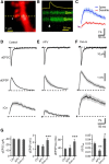
Figure 1.
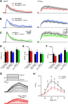
Figure 1.
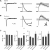
Synaptic potentials and Ca transients evoked at individual dendritic spines of CWCs are enhanced by activation of mAChRs. A, High-magnification image of a spiny region of a dendrite of a CWC neuron filled with 20 μm Alexa Fluor-594 (red). B, Green fluorescence (Fluo-5F, 300 μm) during a line scan, as indicated by the dashed line in A, that intersects the spine head (Spine) and neighboring dendrite (Den) during glutamate uncaging onto the spine head. The circle in A and arrowhead in B indicate the location and timing, respectively, of a 1 ms pulse of 720 nm laser light used to trigger two-photon-mediated photolysis of MNI-glutamate. The increase in green fluorescence indicates increased intracellular Ca. The yellow trace shows the uEPSP recorded simultaneously at the soma. C, Quantification of the green fluorescence in the spine head and dendrite (n = 25 spines/7 cells, dendrite ΔG/Gsat = 0.6 ± 0.06% vs spine ΔG/Gsat = 3.1 ± 0.3%, p < 0.001). In this and all subsequent figures, the solid line and shaded regions depict the average ± SEM, respectively. D–F, uEPSC (top), uEPSP (middle), and ΔGuEPSP/Gsat (bottom) measured, respectively, in control conditions, in the presence of NMDAR antagonist d-APV (50 μm), and in the presence of the mAChR agonist Oxo-m (1 μm). G–J, Summary of uEPSC amplitude, uEPSP amplitude, uEPSP decay time constant, and ΔCaspine, measured in control conditions (Ctrl), d-APV, and Oxo-m. For this and subsequent figures, asterisks indicate a significant difference from control: *p < 0.05, **p < 0.01, ***p < 0.001.
Figure 2.
Figure 2.
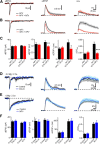
BK channel blockade mimics the mAChR activation effects, whereas SK channels have no effect on uEPSP and Ca signal in CWC spines. A, B, uEPSC (left), uEPSP (middle), and ΔGuEPSP/Gsat (right) measured in the presence of apamin (A) and iberiotoxin (B). The range of amplitudes ± SEM of the uEPSP and ΔGuEPSP/Gsat measured in control conditions are shown by the gray shaded bars. C, Summary of the uEPSC amplitude, uEPSP amplitude, uEPSP decay time constant, and ΔGuEPSP/Gsat in each pharmacological condition.
Figure 3.
Figure 3.
Effects of mAChR activation on synaptic potentials and Ca influx are occluded by blocking BK channels. A, uEPSC, uEPSP, and ΔGuEPSP/Gsat measured in the presence of the antagonists iberiotoxin (IbTx; black) and iberiotoxin plus Oxo-m (red). B, Summary of the uEPSC amplitude, uEPSP amplitude, uEPSP decay time constant, and ΔGuEPSP/Gsat in each condition.
Figure 4.
Figure 4.
NMDAR activity is required for iberiotoxin (IbTx)-induced uEPSP and ΔCaspine increases. A, uEPSC (left), uEPSP (middle), and ΔGuEPSP/Gsat (right) measured in the presence of d-APV (black) and d-APV plus iberiotoxin (red). B, uEPSC (left), uEPSP (middle), and ΔGuEPSP/Gsat (right) measured in the presence of iberiotoxin (black) and iberiotoxin plus d-APV(red). C, Summary plots of uEPSC amplitude, uEPSP amplitude, uEPSP decay time constant, and ΔGuEPSP/Gsat measured as indicated in A and B. D, uEPSC (left), uEPSP (middle), and ΔGuEPSP/Gsat (right) measured in control (black) and Oxo-m (blue) in ACSF containing 0.2 mm magnesium and 2 mm Ca. E, uEPSC (left), uEPSP (middle), and ΔGuEPSP/Gsat (right) measured in control (black) and iberiotoxin (blue) in nominally magnesium-free (0 mm Mg, 2 mm Ca) ACSF. F, Summary plots of amplitudes of uEPSCs, uEPSPs, uEPSP decay time constant, and ΔGuEPSP/Gsat (bottom) measured in the condition shown in D and E.
Figure 5.
Figure 5.
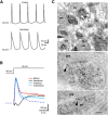
CWCs express rapidly inactivating BK channels. A, BK channel blockade using iberiotoxin reduced the afterhyperpolarization, increasing the tendency to fire complex spikes. B, Depolarizing voltage steps (from −90 to −30 mV, 200 ms) in a CWC elicited a rapidly inactivating outward current that was blocked by iberiotoxin (100 nm). C, Localization of BK antibody labeling in presumptive CWC spines in the DCN using preembedding silver/gold-toned immunoperoxidase (top micrographs) and postembedding immunogold labeling (arrowheads in bottom micrographs). den, Dendrite; pre, presynaptic terminal; sp, spine; asterisk, postsynaptic density. Scale bars: top, 500 nm; bottom, 100 nm.
Figure 6.
Figure 6.
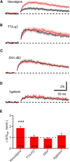
Differential regulation of ΔCauEPSP by voltage-sensitive Ca channels. A–D, ΔCaspine measured in the presence of antagonists of L-type (nimodipine), T-type (TTA-P2), R-type (SNX-482), and P/Q-type (ω-agatoxin IVA) Ca channels, respectively. E, Summary plots of amplitudes of ΔG/Gsat measured in the conditions show in A–D.
Figure 7.
Figure 7.
The effect of mAChR activation and BK channel blockade on synaptic potential and Ca influx are occluded by the L-type Ca channel blocker nimodipine (Nimo). A, uEPSP (left) and ΔCaspine (right) measured in the presence of nimodipine (black) and nimodipine plus iberiotoxin (IbTx) (red). B, uEPSP (left) and ΔCaspine (right) measured in the presence of nimodipine (black) and nimodipine plus Oxo-m (blue). C, uEPSP (left) and ΔCaspine (right) measured in the presence of Bay-K 8644 (black) and Bay-K plus Oxo-m (green). D–F, Summary plots of uEPSP amplitude, uEPSP decay time constant, and ΔCaspine measured in the condition shown in A–C. G, Voltage steps from −70 mV to a potential of −50 to +20 mV (300 ms in 10 mV increments) evoked Ca influx into the spine head in control (black) and Oxo-m (red) through L-type Ca channels (p = 0.03, 0.02, 0.01, 0.04, 0.02 at −40, −30, −20, −10, and 0 mV respectively). H, Summary data for ΔCaspine measured in control conditions and in the presence of Oxo-m.
Figure 8.
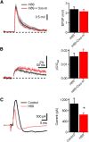
Figure 8.
mAChR activation decreases BK current through PKA signaling. A, Left, uEPSP measured in the presence of H89, a PKA inhibitor (black), and H89 plus Oxo-m (red, left). Right, Bar graph of amplitudes of uEPSP measured in the condition shown in the left. B, Left, ΔCaspine measured in the presence of H89 (black) and H89 plus Oxo-m (red). Right, Bar graphs of ΔCaspine measured in the condition show in the left. C, Left, Traces illustrating BK current extracted by voltage step and iberiotoxin (from −90 to −30 mV) in the presence of control (black) and H89 (red). Right, Bar graph of BK current in the above conditions.
Figure 9.
Figure 9.
Schematic diagram of CWC spine signaling pathways. Glutamate activates AMPARs and NMDARs producing an EPSP and Ca entry through NMDARs. Synaptic stimulation under control conditions also activates L-type Ca channels that contribute to BK channel gating, generating a hyperpolarizing current that suppresses both the EPSP and Ca transient. Activation of mAChRs reduces Ca entry through L-type channels through the PKA signaling pathway, thereby reducing BK activation and enhancing both the EPSP amplitude and Ca influx through NMDARs.

Similar articles

Cited by

References

    1. Bauer CA, Brozoski TJ. Assessing tinnitus and prospective tinnitus therapeutics using a psychophysical animal model. J Assoc Res Otolaryngol. 2001;2:54–64. doi: 10.1007/s101620010030. - DOI - PMC - PubMed
    1. Bender KJ, Uebele VN, Renger JJ, Trussell LO. Control of firing patterns through modulation of axon initial segment T-type calcium channels. J Physiol. 2012;590:109–118. doi: 10.1113/jphysiol.2011.218768. - DOI - PMC - PubMed
    1. Benson TE, Brown MC. Synapses formed by olivocochlear axon branches in the mouse cochlear nucleus. J Comp Neurol. 1990;295:52–70. doi: 10.1002/cne.902950106. - DOI - PubMed
    1. Berkefeld H, Fakler B. Ligand-gating by Ca+ is rate limiting for physiological operation of BK(Ca) channels. J Neurosci. 2013;33:7358–7367. doi: 10.1523/JNEUROSCI.5443-12.2013. - DOI - PMC - PubMed
    1. Bloodgood BL, Sabatini BL. Nonlinear regulation of unitary synaptic signals by CaV(2.3) voltage-sensitive calcium channels located in dendritic spines. Neuron. 2007;53:249–260. doi: 10.1016/j.neuron.2006.12.017. - DOI - PubMed

Publication types

MeSH terms