. 2014 Apr 9;34(15):5322-34.
doi: 10.1523/JNEUROSCI.4703-13.2014.

Activation of spinal glucagon-like peptide-1 receptors specifically suppresses pain hypersensitivity

Affiliations

Activation of spinal glucagon-like peptide-1 receptors specifically suppresses pain hypersensitivity

Nian Gong et al. J Neurosci. .

Abstract

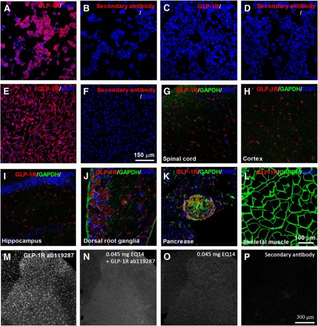
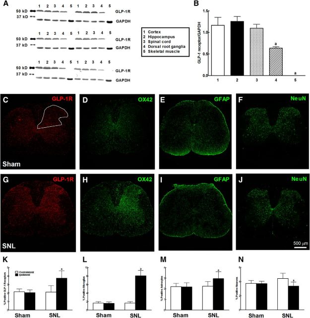
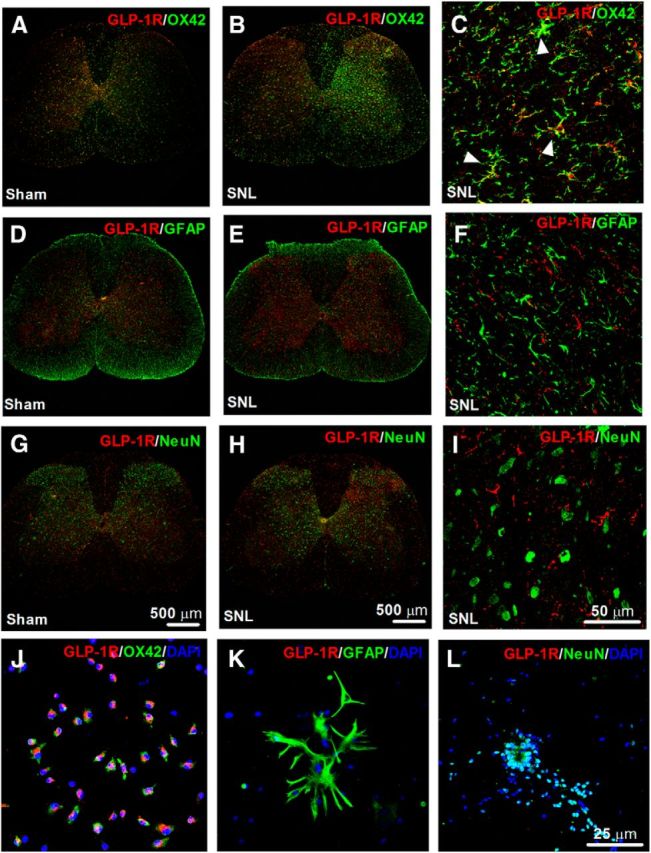
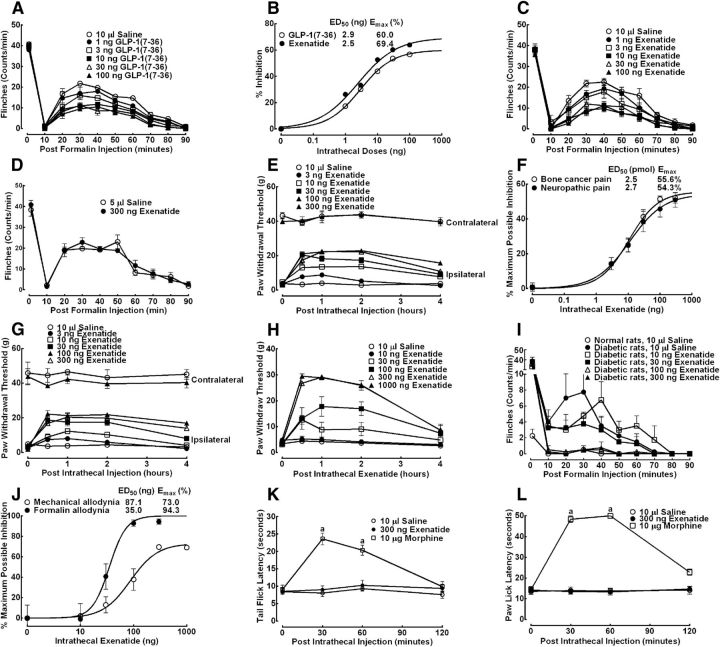
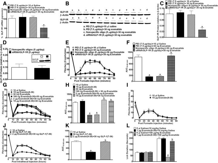
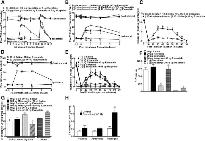
This study aims to identify the inhibitory role of the spinal glucagon like peptide-1 receptor (GLP-1R) signaling in pain hypersensitivity and its mechanism of action in rats and mice. First, GLP-1Rs were identified to be specifically expressed on microglial cells in the spinal dorsal horn, and profoundly upregulated after peripheral nerve injury. In addition, intrathecal GLP-1R agonists GLP-1(7-36) and exenatide potently alleviated formalin-, peripheral nerve injury-, bone cancer-, and diabetes-induced hypersensitivity states by 60-90%, without affecting acute nociceptive responses. The antihypersensitive effects of exenatide and GLP-1 were completely prevented by GLP-1R antagonism and GLP-1R gene knockdown. Furthermore, exenatide evoked β-endorphin release from both the spinal cord and cultured microglia. Exenatide antiallodynia was completely prevented by the microglial inhibitor minocycline, β-endorphin antiserum, and opioid receptor antagonist naloxone. Our results illustrate a novel spinal dorsal horn microglial GLP-1R/β-endorphin inhibitory pathway in a variety of pain hypersensitivity states.

Keywords: GLP-1 receptor; chronic pain; microglia.


Figures

Figure 1.
Figure 1.
Representative photomicrographs of GLP-1R expression in HEK293 cells stably expressing human GLP-1Rs (A, B), HEK293T cells that do not express GLP-1Rs (C, D), PC12 cells expressing rat GLP-1Rs (E, F), and their corresponding negative controls of the secondary antibody (B, D, F), as well as the superficial spinal dorsal horn (laminae I-III; G), cortex (H), hippocampus (I), dorsal root ganglia (J), pancreatic islets (K), and skeletal muscle (L) from 6 normal rats. Tissues were doubly labeled with the GLP-1R and GAPDH antibodies. DAPI staining was also used to determine cell nuclei. Representative photomicrographs for the preventive effect of the GLP-1R antigenic peptide EQ14 on GLP-1R immunoreactive fluorescence staining in the spinal cords from 6 spinal nerve ligation-induced neuropathic rats. M, Specific fluorescence staining with the GLP-1R antibody alone. N, Fluorescence staining with the GLP-1R antibody in the presence of EQ14. O, Fluorescence staining with EQ14 alone. P, Negative control of the secondary antibody. Scale bars: A–F, 150 μm; G–L, 100 μm; M–P, 300 μm.
Figure 2.
Figure 2.
Expression of GLP-1Rs in the spinal cord, cortex, hippocampus, dorsal root ganglia, and skeletal muscle of normal rats by Western blotting where the GAPDH protein was used as loading control: representative gel (A) and GLP-1R/GAPDH band intensity ratio (B). Upregulation of GLP-1R immunostaining specifically expressed on microglia in the spinal dorsal horn after peripheral nerve injury. Photomicrographs were taken from the spinal cords in sham and neuropathic rats. Peripheral neuropathy was induced by unilateral L5–L6 spinal nerve ligation, and frozen sections were obtained from the spinal lumbar enlargements 2 weeks after surgery. Immunofluorescence was labeled with the GLP-1R antibody (C, G), microglial marker CD11b (OX42) (D, H), astrocytic marker GFAP (E, I), and mature neuronal marker NeuN (F, J). GLP-1R-(K) and OX42 (L)/GFAP (M)/NeuN (N)-immunolabeled surface areas were quantified from the spinal dorsal horn (laminae I-V; as indicated in C) using the ImageJ computer program. The averaged percentage immunolabeled surface area was the fraction of the positive immunofluorescent surface area of total measured area in the picture from 3 sections of each spinal cord. Scale bars: 500 μm. Data are mean ± SEM (n = 5 or 6 in each group). aStatistically significant difference from the spinal cord group in B (p < 0.05 by one-way ANOVA followed by post hoc Student-Newman-Keuls test) or the contralateral dorsal horns in K–N (p < 0.05 by paired Student's t test).
Figure 3.
Figure 3.
Representative photomicrographs of GLP-1R double fluorescence labeling with the microglial marker OX42 (A–C), astrocytic marker GFAP (D–F), and mature neuronal marker neuronal nuclei (NeuN; G–I) in the spinal cord. Photomicrographs were taken from the entire spinal cords (A, B, D, E, G, H) and superficial dorsal horns (laminae I-III; C, F, I) in sham and neuropathic rats (n = 5 or 6 in each group), respectively. Peripheral neuropathy was induced by unilateral L5–L6 spinal nerve ligation, and frozen sections were obtained from the spinal lumbar enlargements 2 weeks after surgery. Arrows indicate double-labeling of GLP-1Rs in microglia (C), but not in astrocytes (F) or neurons (I). There is increased fluorescence intensity of labeling of GLP-1R/OX42 in the areas of in the I-V laminae after peripheral nerve injury. Specific expression of GLP-1Rs on all primarily cultured spinal microglial cells (J), but not on astrocytes (K) or neurons (L) from the spinal dorsal horn of neonatal rats. DAPI staining was also used to determine cell nuclei. Scale bars: A, B, D, E, G, H, 500 μm; C, F, I, 50 μm; J–L, 25 μm.
Figure 4.
Figure 4.
Effects of intrathecal and intracerebroventricular single injection of GLP-1(7–36) and exenatide on formalin-induced flinching response (A–D), bone cancer-induced mechanical allodynia (E, F), spinal nerve ligation-induced mechanical allodynia (F, G), diabetes-induced mechanical allodynia (H, J), and formalin-induced flinching response (I, J), and thermally evoked nociceptive reflex responses in the tail immersion test (K) and hotplate test (L) in rats. Dose–response analyses of intrathecal injection of GLP-1(7–36) and exenatide on formalin-induced tonic flinching response (B), bone cancer- and nerve injury-induced mechanical allodynia (F), and diabetic mechanical and formalin-induced flinching response (J), best fitted by the nonlinear least-squares method. For the formalin test, naive and diabetic rats (∼30–50 d after intravenous injection of 50 mg/kg streptozocin) received intrathecal injection of saline, GLP-1(7–36), or exenatide 30 min before paw injection of 50 μl of 5% or 0.2% formalin, respectively. Nociceptive behavior was quantified by counting the number of formalin-injected paw flinches in 1 min epochs. For the bone cancer-, peripheral nerve injury-, and diabetes-induced mechanical allodynia, the paw withdrawal thresholds were measured by electronic von Frey filaments ∼14–50 d after tibia implantation of Walker 256 cancer cells, tight ligation of L5–L6 spinal nerves, and intravenous injection of streptozocin (50 mg/kg), respectively. Data are mean ± SEM (n = 6 in each group). aStatistical significance compared with the saline control group (p < 0.05 by two-way ANOVA followed by post hoc Student-Newman-Keuls test).
Figure 5.
Figure 5.
Effects of 7 day intrathecal injections of the GLP-1R gene silencer siRNA/GLP-1R on spinal GLP-1R gene expression (relative to gapdh) (A), spinal (B, C), and dorsal root ganglial (D, with representative gels as in the inset) GLP-1R protein expression (relative to β-actin) and formalin-induced flinching responses (E, F) in rats. The vehicle PEI (7.5 μg) control group, nonspecific oligoneucleotides (oligo, 5 μg) control group, and siRNA/GLP-1R (5 μg) group were multidaily intrathecally injected for 7 d in rats. On the eight day, rats received a single intrathecal injection of saline (10 μl) or exenatide (30 ng) before 5% formalin challenge. Nociceptive behavior was quantified by counting the number of paw flinches in 1 min epochs. For the GLP-1R expression study, homogenates were obtained from spinal lumbar enlargements or dorsal rood ganglia immediately after the completion of the behavior tests. Blockade effects of intrathecal injection of the specific GLP-1R antagonist exendin(9–39) on antinociceptive effects of GLP-1(7–36) and exenatide in the rat and mouse formalin test (G–L). Rats and mice received two intrathecal treatments 30 min before subcutaneous injection of 10 or 50 μl of 1% or 5% formalin. The cumulated licking/biting duration from 0 to 5 min and 20 to 40 min in mice after formalin injection represented acute nociception and tonic hyperalgesia, respectively. Data are mean ± SEM (n = 6 in each group). a,bStatistical significance compared with the vehicle control and the exenatide or GLP-1(7–36) group, respectively (p < 0.05 by one-way ANOVA followed by post hoc Student-Newman-Keuls test).
Figure 6.
Figure 6.
Effects of intrathecal injection of the microglial inhibitor minocycline (A), specific β-endorphin antiserum (B, C), and specific opioid receptor antagonist naloxone (D–F) on spinal exenatide antinociception in peripheral neuropathy-induced mechanical allodynia or formalin-induced hyperalgesia in rats. For neuropathic rats, paw withdrawal thresholds were measured by electronic von Frey filaments 2 weeks after tight ligation of L5–L6 spinal nerves. For the formalin test, rats received two intrathecal treatments 30 min before subcutaneous injection of 50 μl of 5% formalin. Nociceptive behavior was quantified by counting the number of the formalin-injected paw flinches in 1 min epochs. Blank serum as the negative control was from a healthy rabbit without any treatment. G, Effects of intrathecal injection of exenatide on the spinal β-endorphin level in neuropathic rats and sham rats. Minocycline (100 μg) was intrathecally injected 4 h earlier before exenatide treatment. Ipsilateral spinal lumbar enlargements were obtained 1 h after exenatide injection in neuropathic rats. H, Effects of exenatide (10−8 m) on β-endorphin release at 2 h after application in primarily cultured spinal microglia, astrocytes, and neurons from the spinal dorsal horn of neonatal rats. β-Endorphin levels in the spinal cord homogenates and culture media were determined by a specific fluorescent immunoassay kit. Data are means ± SEM (n = 6 in each group). a,bStatistical significance compared with the saline control and the exenatide group, respectively (p < 0.05 by one-way or two-way ANOVA followed by post hoc Student-Newman-Keuls test).cStatistical significance compared with the saline control in sham rats (p < 0.05 by one- or two-way ANOVA followed by post hoc Student-Newman-Keuls test).

Similar articles

Cited by

References

    1. Alvarez E, Martínez MD, Roncero I, Chowen JA, García-Cuartero B, Gispert JD, Sanz C, Vázquez P, Maldonado A, de Cáceres J, Desco M, Pozo MA, Blázquez E. The expression of GLP-1 receptor mRNA and protein allows the effect of GLP-1 on glucose metabolism in the human hypothalamus and brainstem. J Neurochem. 2005;92:798–806. doi: 10.1111/j.1471-4159.2004.02914.x. - DOI - PubMed
    1. Avila-Martin G, Galan-Arriero I, Gómez-Soriano J, Taylor J. Treatment of rat spinal cord injury with the neurotrophic factor albumin-oleic acid: translational application for paralysis, spasticity and pain. PLoS One. 2011;6:e26107. doi: 10.1371/journal.pone.0026107. - DOI - PMC - PubMed
    1. Bach FW. Beta-endorphin in the brain: a role in nociception. Acta Anaesthesiol Scand. 1997;41:133–140. doi: 10.1111/j.1399-6576.1997.tb04627.x. - DOI - PubMed
    1. Baggio LL, Drucker DJ. Biology of incretins: GLP-1 and GIP. Gastroenterology. 2007;132:2131–2157. doi: 10.1053/j.gastro.2007.03.054. - DOI - PubMed
    1. Beinborn M, Worrall CI, McBride EW, Kopin AS. A human glucagon-like peptide-1 receptor polymorphism results in reduced agonist responsiveness. Regul Pept. 2005;130:1–6. doi: 10.1016/j.regpep.2005.05.001. - DOI - PubMed

Publication types

MeSH terms

LinkOut - more resources