Review
. 2014 Mar-Apr;3(2):179-91.
doi: 10.1002/wdev.128. Epub 2013 Oct 16.

The Drosophila auditory system

Affiliations
Review

The Drosophila auditory system

Grace Boekhoff-Falk et al. Wiley Interdiscip Rev Dev Biol. 2014 Mar-Apr.

Abstract

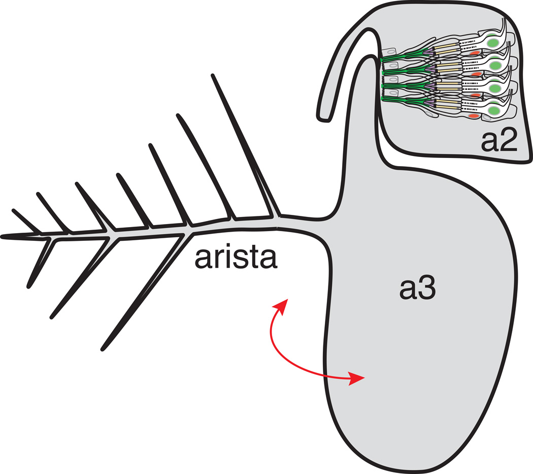
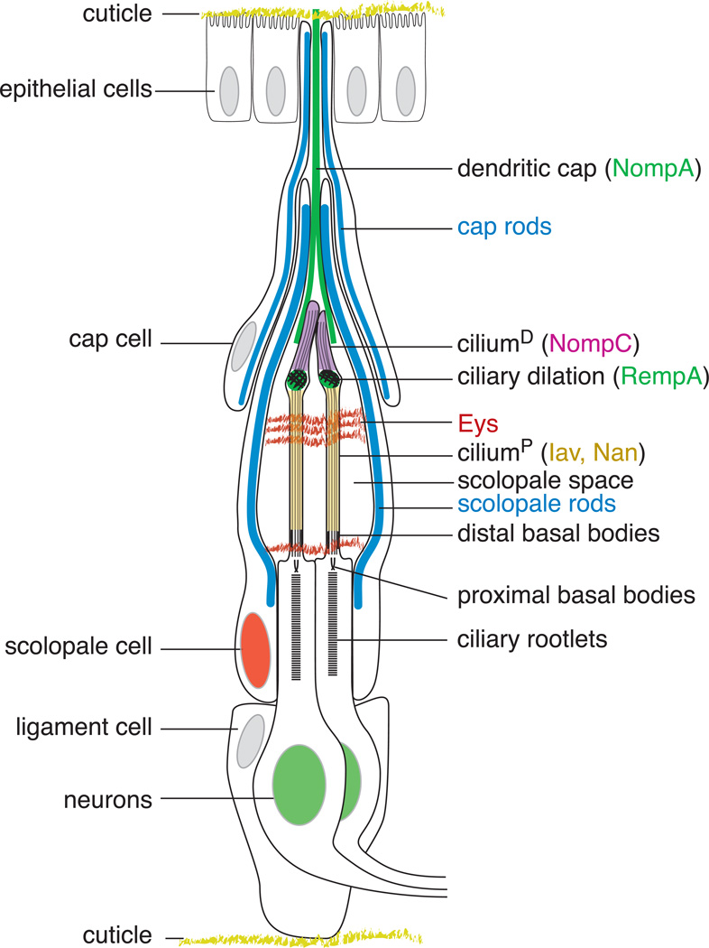
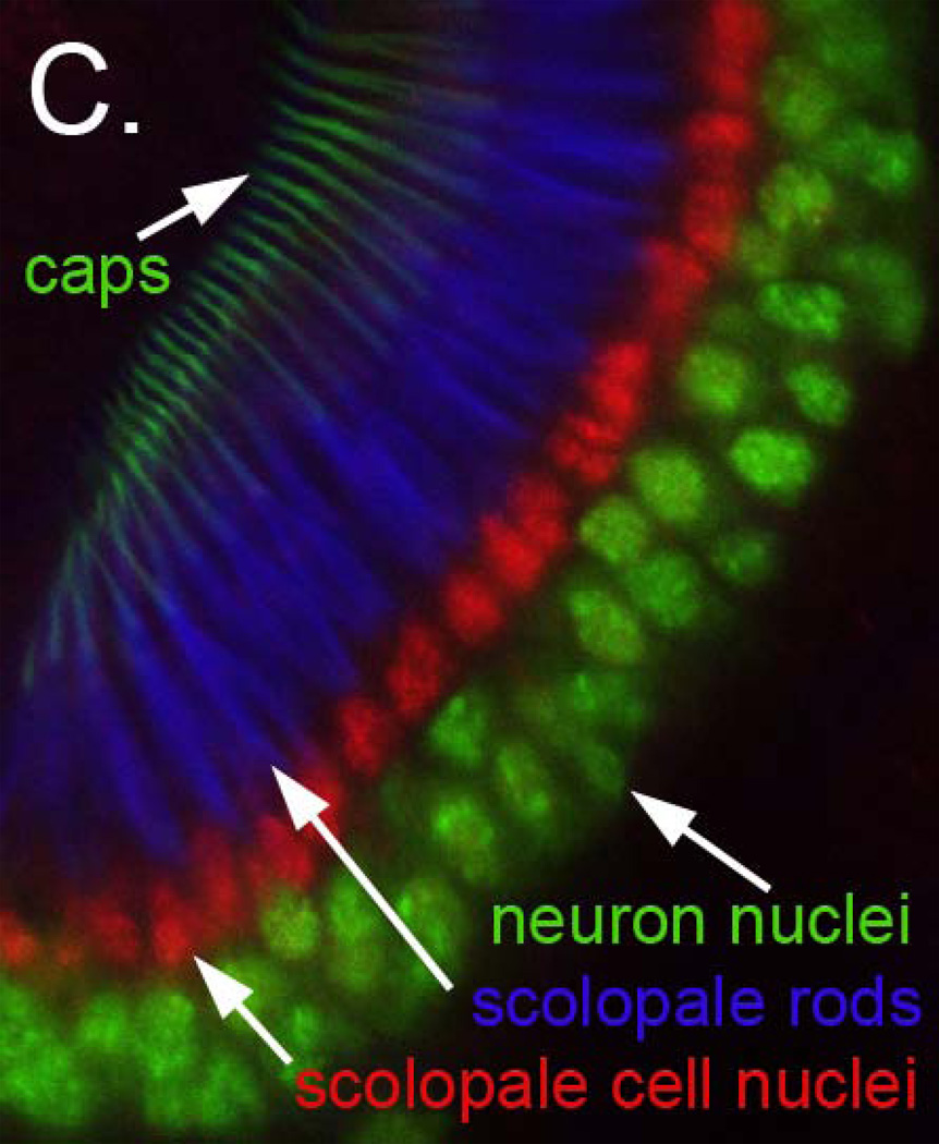
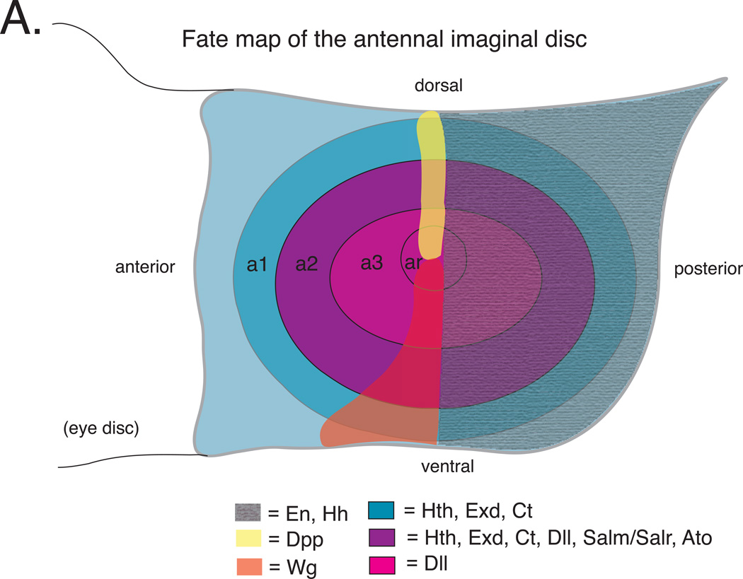
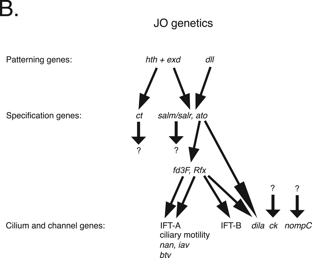
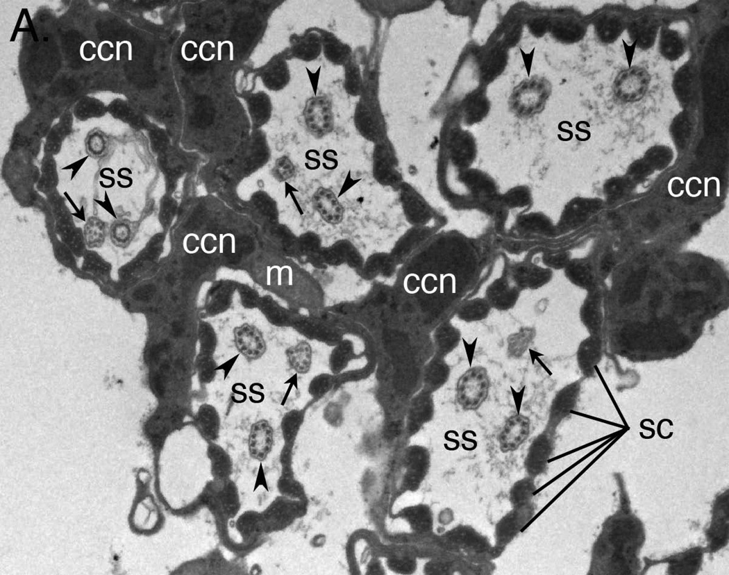
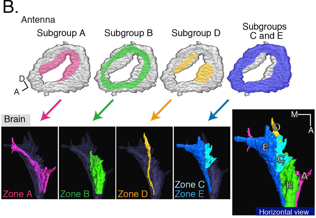
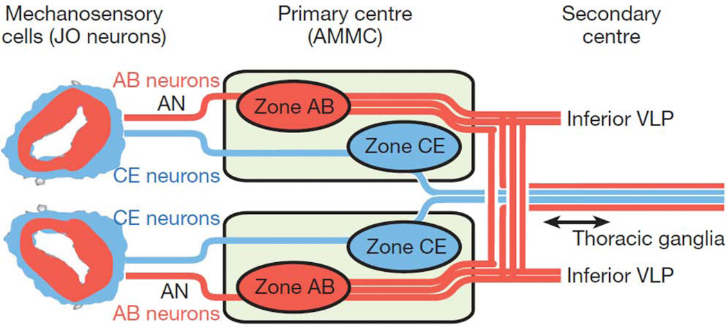
Development of a functional auditory system in Drosophila requires specification and differentiation of the chordotonal sensilla of Johnston's organ (JO) in the antenna, correct axonal targeting to the antennal mechanosensory and motor center in the brain, and synaptic connections to neurons in the downstream circuit. Chordotonal development in JO is functionally complicated by structural, molecular, and functional diversity that is not yet fully understood, and construction of the auditory neural circuitry is only beginning to unfold. Here, we describe our current understanding of developmental and molecular mechanisms that generate the exquisite functions of the Drosophila auditory system, emphasizing recent progress and highlighting important new questions arising from research on this remarkable sensory system.


Conflict of interest statement

Grace Boekhoff-Falk, No conflict of interest.

Daniel F. Eberl, No conflict of interest.

Figures

Figure 1
Figure 1. Johnston’s organ develops in the second antennal segment
A. Schematic of the adult Drosophila antenna in which Johnston’s organ resides. Sound displaces the arista, rotating (red arrow) the olfactory third antennal segment (a3). Johnston’s organ in the second antennal segment (a2) serves as the mechanoreceptor for hearing, and also responds to antennal deflections induced by wind or gravity. Only four of more than 225 scolopidia are depicted here, and an individual scolopidium is depicted in greater detail in Figure 1B. B. Schematic of a typical Drosophila Johnston’s organ scolopidium. The major structural elements of JO are diagrammed. The scolopidia are suspended between the cuticle attachments at a2/a3 joint (top) and the peripheral cuticle of a2 (bottom), with the dendritic cap (marked by the NompA protein (green)) and ligament cells forming the respective connections. The cap rods and scolopale rods, made of thick actin filaments surrounding nucleating microtubules, are shown in blue. The scolopale cell wraps the sensory dendrites to form the scolopale space, a tightly sealed extracellular cavity thought to contain a specialized receptor lymph. The Eyes shut (Eys) protein recognized by the 21A6 monoclonal Ab (red) forms an extracellular matrix in the scolopale space, protecting against desiccation at higher temperatures. In the sensory dendrite, the ciliary dilation, a feature unique to chordotonal cilia and marked by the RempA protein (green), delimits the distal cilium (ciliumD), where the TRPN channel NompC is localized (magenta), from the proximal cilium (ciliumP), where the TRPV channel Iav/Nan heteromultimer is localized (yellow). The dendritic cap is drawn proportionately shorter to allow detailed depiction of the scolopale space and sensory cilia. Features shown in Figure 1C are depicting here in matching colors. C. Organization of Johnston’s organ scolopidia. Confocal micrograph of a wild type pupal JO in which neuronal nuclei are labeled in green using an antibody to Embryonic lethal, abnormal vision (Elav), scolopale cell nuclei are labeled in red using an antibody to Prospero (Pros), the actin-rich scolopale rods are labeled in blue using Alexa-633 conjugated phalloidin, and the cap structures are labeled using a NompA-GFP transgene. These features are depicted in panel 1C in matching colors.
Figure 2
Figure 2. Genetics of Johnston’s organ development
A. Schematic of a third instar larval antennal imaginal disc. a1, a2 and a3 = first, second and third antennal segment precursors, respectively. ar = arista precursors. En and Hh are expressed throughout the posterior compartment of the disc. hth and dll are regulated by Dpp and Wg. Hth and Dll expression overlap in presumptive a2 where they activate salm/salr and ato. ato is required for specification of JO precursors. Based on information in references –, , , . B. Johnston’s organ development is controlled by a genetic cascade initiated by transcription factors encoded by hth, exd and Dll. Hth and Exd together activate the expression of ct, while Hth, Exd and Dll together activate salm/salr and ato. Both ct and salm/salr mutants are deaf, exhibiting defective JO development followed by JO degeneration. However, genes regulated by the Ct and salm/salr transcription factors are unknown. Ato directly regulates Rfx and dila expression and either directly or indirectly regulates fd3F expression. Together, the Rfx and Fd3F transcription factors activate the expression of a suite of genes required for ciliogenesis, ciliary motility and JO function, including multiple intraflagellar transport A (IFT-A) genes required for retrograde transport, axonemal dyneins required for ciliary motility, the TRPV channels encoded by iav and nan, and the retrograde IFT dynein motor encoded by btv. In addition, Rfx, but not Fd3F, activates a subset of the IFT-B genes required for anterograde transport. Regulators of the myosin VIIA homolog encoded by ck and the TRPN channel encoded by nompC remain unknown. Not indicated here is the restriction of gene expression to subsets of cell types; whereas patterning genes are expressed throughout presumptive a2, the genes at the bottom of the hierarchy tend to be restricted to either neurons or specific subsets support cells. Note that although Rfx and fd3F are expressed in JO, the targets indicated here were identified in larval chordotonal organs. All of the genes shown here have vertebrate homologs, and most of these also are required for vertebrate ear development and/or function. Based on information in references , , , .
Figure 3
Figure 3. The molecular and structural diversity of Johnston’s organ neurons
A. Transmission electron micrograph showing cross sections of five JO scolopidia. Each scolopidium possesses two dendrites with typical ciliary 9 × 2+0 axonemes (arrowheads). Four of the five scolopidia in this view show a third dendrite (arrows) with a degenerate axoneme or even disordered microtubules. ss = scolopale space; ccn = cap cell nucleus; sc = scolopales; m = mitochondrion. B. Functional diversity of JO neurons. Promoter fusions and enhancer trap lines have been identified that mark subsets of JO neurons. These neurons have been classified into five groups, A–E (upper panels), that innervate distinct zones in the antennal mechanosensory and motor center (AMMC; lower panels). The positions of the Type A neuronal cell bodies within JO are highlighted in pink. The locations of the Type B neuronal cell bodies are indicated in green; the locations of the Type D neuronal cell bodies are highlighted in yellow; and the locations of the Types C and E neuronal cell bodies are indicated in blue. Neuronal types A and B are used for sound reception; types C and E are used for gravity and wind reception; the function of type D neurons remains unknown. In addition to the AMMC, auditory information from some Type A neurons is carried to either the subesophageal ganglion (SOG) or the ventrolateral protocerebrum (vlpr). The SOG also receives gustatory information, while the vlpr also receives visual and olfactory information, suggesting that there is convergence of multiple sensory modalities in these brain regions. Reproduced with kind permission from Springer Science and Business Media .
Figure 4
Figure 4. Drosophila auditory circuitry
From JO, auditory information is relayed to a different region of the antennal mechanosensory and motor complex (AMMC) than wind and gravity information. The auditory neurons and their axon tracts are shown in red, while the wind and gravity sensing neurons and their axon tracts are indicated in blue. Reprinted by permission from Macmillan Publishers Ltd: Nature 458: 165–171 (2009).

References

    1. Hoy RR. Acute as a bug's ear: an informal discussion of hearing in insects. In: Hoy RR, Popper AN, Fay RR, editors. Comparative Hearing: Insects. Vol. 10. New York: Springer; 1998. pp. 1–17.
    1. Eberl DF. Feeling the vibes: chordotonal mechanisms in insect hearing. Curr. Opin. Neurobiol. 1999;9:389–393. - PubMed
    1. Field LH, Matheson T. Chordotonal organs of insects. In: Evans PD, editor. Adv. Insect Physiol. Vol. 27. San Diego: Academic Press; 1998. pp. 1–228.
    1. Todi SV, Franke JD, Kiehart DP, Eberl DF. Myosin VIIA defects, which underlie the Usher 1B Syndrome in humans, lead to deafness. Drosophila. Curr. Biol. 2005;15:862–868. doi: - PMC - PubMed
    1. Todi SV, Sivan-Loukianova E, Jacobs JS, Kiehart DP, Eberl DF. Myosin VIIA, important for human auditory function, is necessary for Drosophila auditory organ development. PLoS ONE. 2008;3:e2115. - PMC - PubMed

Publication types

MeSH terms

LinkOut - more resources