. 2014 Aug;66(8):2188-200.
doi: 10.1002/art.38656.

Peripheral calcitonin gene-related peptide receptor activation and mechanical sensitization of the joint in rat models of osteoarthritis pain

Affiliations
Free PMC article

Peripheral calcitonin gene-related peptide receptor activation and mechanical sensitization of the joint in rat models of osteoarthritis pain

Craig M Bullock et al. Arthritis Rheumatol. 2014 Aug.
Free PMC article

Abstract

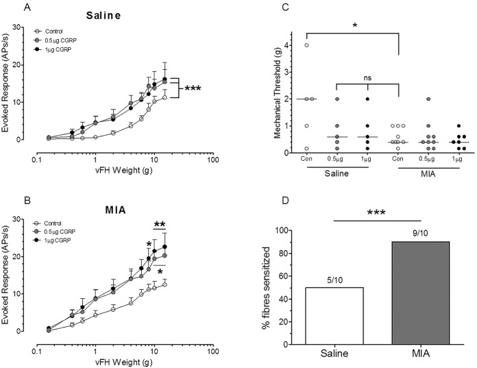
Objective: To investigate the role of the sensory neuropeptide calcitonin gene-related peptide (CGRP) in peripheral sensitization in experimental models of osteoarthritis (OA) pain.

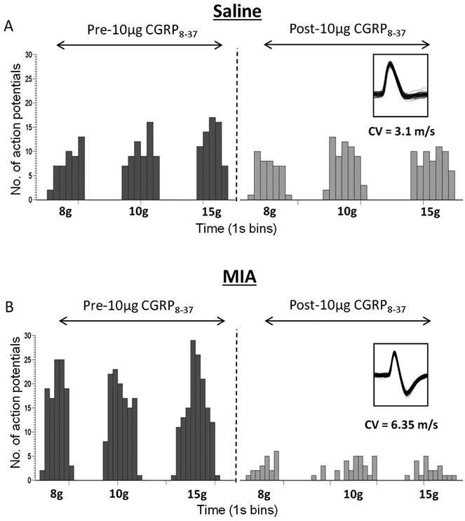
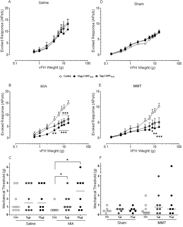
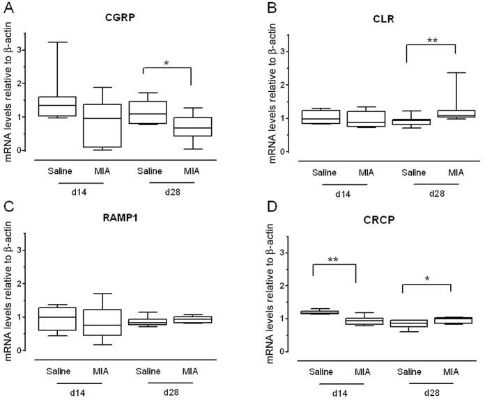
Methods: Experimental knee OA was induced in rats by intraarticular injection of monosodium iodoacetate (MIA) or by transection of the medial meniscus (MMT). Single-unit recordings of joint-innervating nociceptors were obtained in MIA- and saline-treated rats following administration of CGRP or the CGRP receptor antagonist CGRP 8-37. Effects of CGRP 8-37 were also examined in rats that underwent MMT and sham operations. Protein and messenger RNA (mRNA) levels of CGRP receptor components in the L3-L4 dorsal root ganglion (DRG) were investigated following MIA treatment.

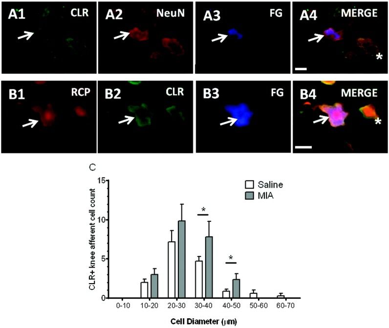
Results: In both the MIA and MMT groups, the mechanical sensitivity of joint nociceptors was enhanced compared to that in the control groups. Exogenous CGRP increased mechanical sensitivity in a greater proportion of joint nociceptors in the MIA-treated rats than in the saline-treated rats. Local blockade of endogenous CGRP by CGRP 8-37 reversed both the MIA- and MMT-induced enhancement of joint nociceptor responses. Joint afferent cell bodies coexpressed the receptor for CGRP, called the calcitonin-like receptor (CLR), and the intracellular accessory CGRP receptor component protein. MIA treatment increased the levels of mRNA for CLR in the L3-L4 DRG and the levels of CLR protein in medium and large joint afferent neurons.

Conclusion: Our findings provide new and compelling evidence implicating a role of CGRP in peripheral sensitization in experimental OA. Our novel finding of CGRP-mediated control of joint nociceptor mechanosensitivity suggests that the CGRP receptor system may be an important target for the modulation of pain during OA. CGRP receptor antagonists recently developed for migraine pain should be investigated for their efficacy against pain in OA.


Figures

Figure 1
Figure 1
Calcitonin gene-related peptide (CGRP)–induced mechanical sensitization of an increased proportion of knee afferents following monosodium iodoacetate (MIA) injection. A and B, CGRP significantly increased the mechanical evoked firing rates (action potentials per second [APs/s]) of joint afferents in saline-treated (A) and MIA-treated (B) rats (P < 0.001 by 2-way analysis of variance with Bonferroni post hoc test). Values are the mean ± SEM maximum sensitizing effects of CGRP over each 1-hour recording period (n = 9 MIA-treated rats; n = 5 saline-treated rats). C, CGRP administration reduced mechanical thresholds (minimum stimulus evoking ≥2 action potentials) in saline-treated rats to a level not significantly different from that in MIA-treated rats (P > 0.05 for comparisons of 0.5 μg or 10 μg of CGRP versus controls, by Mann-Whitney t-test). Mechanical thresholds in MIA-treated rats were unaltered by CGRP administration. Each circle represents an individual rat (n = 7–9 MIA treated and n = 5 saline treated); horizontal lines show the median. D, The percentage of fibers sensitized by CGRP was greater in MIA-treated rats than in saline-treated rats (P < 0.001 by Fisher's exact test). ∗ = P < 0.05; ∗∗ = P < 0.01; ∗∗∗ = P < 0.001 by Mann-Whitney t-test. vFH = von Frey hair; NS = not significant.
Figure 2
Figure 2
Inhibition of knee afferent mechanical evoked firing by the calcitonin gene-related peptide (CGRP) receptor antagonist CGRP 8–37 in monosodium iodoacetate (MIA)–treated rats. Representative examples of knee afferent mechanical evoked (8-, 10-, or 15-gm von Frey monofilaments) response data recorded before and after administration of 10 μg of CGRP 8–37 in a saline-treated rat (A) and an MIA-treated rat (B) are shown. The inhibitory effects of CGRP 8–37 were more pronounced following MIA treatment. Results are represented as rate histograms (1-second bins) of action potential firing of knee afferents (single units) in response to stimulation at the indicated von Frey monofilament level. Insets show an overlay of a representative fiber recording with the corresponding conduction velocity (CV; in meters/second), demonstrating single-fiber recordings; both are Aδ fibers.
Figure 3
Figure 3
Ablation of monosodium iodoacetate (MIA)–induced and medial meniscus transection (MMT)–induced mechanical sensitization of joint afferents by the calcitonin gene-related peptide (CGRP) receptor antagonist CGRP 8–37. CGRP 8–37 had no effect on mechanically evoked responses (action potentials per second [APs/s]) of joint afferents in saline-treated (A) and sham-operated (D) rats, whereas it significantly inhibited mechanically evoked responses in MIA-treated (B) and MMT-operated (E) rats (P < 0.0001 for each comparison versus its corresponding control, by 2-way analysis of variance with Bonferroni post hoc test). Values are the mean ± SEM responses over each 1-hour recording period (n = 8 saline-treated, 6 sham-operated, 9 MIA-treated, and 8 MMT-operated rats). CGRP 8–37 had no effect on mechanical thresholds (minimum stimulus evoking ≥2 action potentials) in saline-treated (C) or sham-operated (F) rats. However, in MIA-treated (C) and MMT-operated (F) rats, thresholds in the presence of 10 μg of CGRP 8–37 were increased, reversing the osteoarthritis-induced reduction of mechanical thresholds. Each circle represents an individual rat (n = 6–9 per group); horizontal lines show the median. ∗ = P < 0.05; ∗∗ = P < 0.01; ∗∗∗ = P < 0.001, by Mann-Whitney t-test. vFH = von Frey hair.
Figure 4
Figure 4
Expression of mRNA for calcitonin gene-related peptide (CGRP) receptor components in knee joint–innervating L3–L4 dorsal root ganglia (DRGs) and modulation of the levels following treatment with monosodium iodoacetate (MIA). A, Expression of mRNA for CGRP was significantly reduced 28 days after MIA treatment. B, Calcitonin-like receptor (CLR) expression was increased 28 days after MIA treatment. C, Levels of receptor activity–modifying protein 1 (RAMP-1) were unaltered, both on day 14 and on day 28 after MIA treatment. D, Levels of mRNA for CGRP receptor component protein (CRCP) were reduced 14 days after MIA treatment. There was a small but significant increase in CRCP expression 28 days after MIA treatment. Results were normalized to β-actin expression (which was unchanged between groups and treatments). Data are shown as box plots. Each box represents the 25th to 75th percentiles (n = 6–8 rats per group). Lines inside the boxes represent the median. Lines outside the boxes represent the 10th and 90th percentiles. ∗ = P < 0.05; ∗∗ = P < 0.01 by Mann-Whitney t-test.
Figure 5
Figure 5
Coexpression of the calcitonin gene-related peptide (CGRP) receptor components calcitonin-like receptor (CLR) and receptor component protein (CRCP [RCP]) by knee afferent neurons and increased CLR expression following treatment with monosodium iodoacetate (MIA). A1–A4, CLR is expressed on knee afferents (arrows) and non–knee afferent neuronal cell bodies (∗) in ipsilateral L4 dorsal root ganglia (DRGs). Slides incubated with anti–neuronal nuclei (anti-NeuN) antibodies or with Fluoro-Gold (FG) are also shown. Bar = 34 μm. B1–B4, CRCP is coexpressed with CLR on knee afferent neurons (arrows) and non–knee afferent neurons (∗). Bar = 40 μm. C, The numbers of medium (30–40 μm in diameter) and large (40–50 μm in diameter) CLR-positive knee afferents were significantly increased in MIA-treated rats. CLR-positive knee afferent cell counts were analyzed according to diameter ranges similar to those described in a previously published report (9). Values are the mean ± SEM of 7 rats per group. ∗ = P < 0.05 by Mann-Whitney t-test.

Similar articles

Cited by

References

    1. Dieppe PA, Lohmander LS. Pathogenesis and management of pain in osteoarthritis. Lancet. 2005;365:965–73. - PubMed
    1. Kosek E, Ordeberg G. Lack of pressure pain modulation by heterotopic noxious conditioning stimulation in patients with painful osteoarthritis before, but not following, surgical pain relief. Pain. 2000;88:69–78. - PubMed
    1. Creamer P, Hunt M, Dieppe P. Pain mechanisms in osteoarthritis of the knee: effect of intraarticular anesthetic. J Rheumatol. 1996;23:1031–6. - PubMed
    1. Dye SF, Vaupel GL, Dye CC. Conscious neurosensory mapping of the internal structures of the human knee without intraarticular anesthesia. Am J Sports Med. 1998;26:773–7. - PubMed
    1. Arendt-Nielsen L, Nie H, Laursen MB, Laursen BS, Madeleine P, Simonsen OH, et al. Sensitization in patients with painful knee osteoarthritis. Pain. 2010;149:573–81. - PubMed

Publication types

Substances