. 2014 May 23;289(21):14996-5004.
doi: 10.1074/jbc.M113.533547. Epub 2014 Apr 9.

The protein phosphatase 2A regulatory subunit B56γ mediates suppression of T cell receptor (TCR)-induced nuclear factor-κB (NF-κB) activity

Affiliations

The protein phosphatase 2A regulatory subunit B56γ mediates suppression of T cell receptor (TCR)-induced nuclear factor-κB (NF-κB) activity

Rebecca Breuer et al. J Biol Chem. .

Abstract

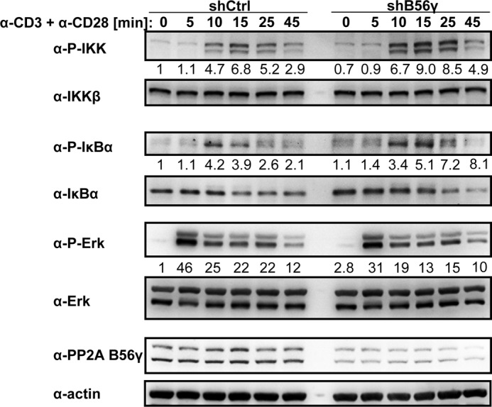
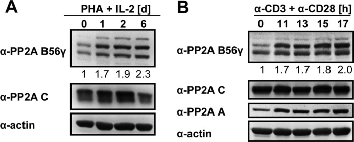
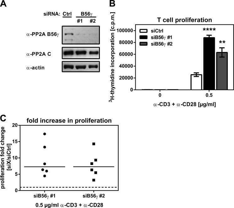
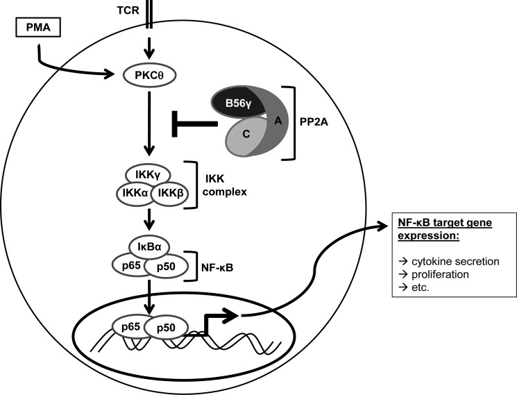
NF-κB is an important transcription factor in the immune system, and aberrant NF-κB activity contributes to malignant diseases and autoimmunity. In T cells, NF-κB is activated upon TCR stimulation, and signal transduction to NF-κB activation is triggered by a cascade of phosphorylation events. However, fine-tuning and termination of TCR signaling are only partially understood. Phosphatases oppose the role of kinases by removing phosphate moieties. The catalytic activity of the protein phosphatase PP2A has been implicated in the regulation of NF-κB. PP2A acts in trimeric complexes in which the catalytic subunit is promiscuous and the regulatory subunit confers substrate specificity. To understand and eventually target NF-κB-specific PP2A functions it is essential to define the regulatory PP2A subunit involved. So far, the regulatory PP2A subunit that mediates NF-κB suppression in T cells remained undefined. By performing a siRNA screen in Jurkat T cells harboring a NF-κB-responsive luciferase reporter, we identified the PP2A regulatory subunit B56γ as negative regulator of NF-κB in TCR signaling. B56γ was strongly up-regulated upon primary human T cell activation, and B56γ silencing induced increased IκB kinase (IKK) and IκBα phosphorylation upon TCR stimulation. B56γ silencing enhanced NF-κB activity, resulting in increased NF-κB target gene expression including the T cell cytokine IL-2. In addition, T cell proliferation was increased upon B56γ silencing. These data help to understand the physiology of PP2A function in T cells and the pathophysiology of diseases involving PP2A and NF-κB.

Keywords: B56γ; IL-2; NF-κB Transcription Factor; Nuclear Factor-κB (NF-κB); PPP2R5C; Phosphoprotein Phosphatase; Serine/Threonine Protein Phosphatase; Signaling; T Cell; T Cell Receptor.


Figures

FIGURE 1.
FIGURE 1.
RNAi screen identifies B56γ (gene name: PPP2R5C) as negative regulator of TCR-mediated NF-κB activation. GLuc-J16 T cells were transfected with a commercially available siRNA library targeting all known or predicted phosphatase genes. Three independent siRNAs (siRNA 1–3) per gene were transfected in duplicate. 72 h after transfection, cells were stimulated for 5 h via the TCR with α-CD3 + α-CD28 antibodies, and supernatants with secreted Gaussia luciferase were taken to measure NF-κB activity. Values were normalized to cell viability with CellTiter-Glo. For a detailed description see Ref. and “Experimental Procedures.” Shown is a reanalysis of siRNA screen data from Ref. . A, decision tree for candidate selection from siRNA screen. B, Z scores for all siRNAs targeting regulatory PP2A subunits and for replicate transfections separately (replicates 1 and 2). Z scores higher than +1.5 are highlighted in dark gray. Z scores between +1 and +1.5 are marked in light gray.
FIGURE 2.
FIGURE 2.
B56γ suppresses NF-κB activity upon TCR, TNFα, and PMA stimulation. A and B, GLuc-J16 T cells were transfected with nontargeting siRNA (siCtrl) and two independent siRNA oligonucleotides targeting B56γ (siB56γ #1 and #2). After 72 h, B56γ knockdown efficiency was assessed by qPCR (A), and cells were stimulated via the TCR for 5 h with α-CD3 + α-CD28 antibodies or left unstimulated (B). NF-κB activity was measured with a Gaussia luciferase assay. Values were normalized to cell viability using CellTiter-Glo. C–F, Jurkat T cells were transfected with an expression plasmid encoding HA-tagged B56γ or empty vector control. In addition, a NF-κB reporter system was cotransfected to determine NF-κB activity. 48 h after transfection B56γ overexpression was confirmed by Western blotting (C), and cells were stimulated as indicated for 5 h (D–F). NF-κB activity was measured using a reporter gene assay. **, p < 0.01; ***, p < 0.001; ****, p < 0.0001. Error bars, S.D.
FIGURE 3.
FIGURE 3.
B56γ suppresses TCR-induced IKK and IκBα phosphorylation. Jurkat T cells were stably transduced with lentiviral particles containing inducible B56γ shRNA or shRNA control. Transduced cells were selected with puromycin. Inducible B56γ knockdown cells and control cells were treated with doxycycline to induce shRNA expression. After 3 days, cells were stimulated via the TCR with 0.5 μg/ml α-CD3 + α-CD28 antibodies for the indicated time points. Lysates were analyzed using Western blotting. Bands were quantified, and the phospho signal was normalized to the respective total protein signal. The siCtrl 0 time point was set to 1. Numbers below the panels indicate relative quantification of signal intensity. Results are representative of two independent experiments.
FIGURE 4.
FIGURE 4.
B56γ expression is up-regulated upon T cell stimulation. A, primary human T cells were stimulated for 24 h with PHA and expanded in the presence of exogenous IL-2 for up to 6 days. Samples were taken at the indicated time points, and protein lysates were analyzed by Western blotting. Bands were quantified, and the B56γ signal was normalized to the respective actin signal. The 0 time point was set to 1. Numbers below the panels indicate relative quantification of signal intensity. One representative of five donors is shown. B, primary human T cells were stimulated via the TCR with α-CD3 + α-CD28 antibodies for the indicated time points. Lysates were analyzed by Western blotting. Bands were quantified, and the B56γ signal was normalized to the respective actin signal. The 0 time point was set to 1. Numbers below the panels indicate relative quantification of signal intensity. One representative of three donors is shown.
FIGURE 5.
FIGURE 5.
B56γ suppresses NF-κB target gene transcription. Primary human T cells were activated with PHA and IL-2 for 3 days. Afterward cells were transfected with siRNA targeting B56γ (siB56γ) or nontargeting siRNA control (siCtrl). A, after 72 h B56γ knockdown was confirmed by Western blotting. B, 72 h after transfection cells were stimulated with 0.5 μg/ml α-CD3 + α-CD28 antibodies for the indicated time points. cDNA of cells was analyzed in a Taqman-based custom designed qPCR array. Data were normalized to three housekeeping gene controls (GAPDH, β-actin, and 18S RNA). Values for nontargeting siRNA at 60 min of stimulation were set to 1. C, 72 h after transfection cells were stimulated with 0.5 μg/ml α-CD3 + α-CD28 antibodies for the indicated time points. cDNA of cells was analyzed for IL-2 transcription in a SYBR Green-based qPCR assay. Values represent mean, and error bars represent maximum and minimum.
FIGURE 6.
FIGURE 6.
B56γ suppresses TCR-induced IL-2 secretion. Primary human T cells were activated with PHA and IL-2 for 3 days. Afterward cells were transfected with nontargeting siRNA (Ctrl), two independent siRNA oligonucleotides targeting B56γ or siRNA targeting the known NF-κB negative regulator CYLD. A, 72 h after transfection, knockdown was confirmed by Western blotting. B, 72 h after transfection, cells were stimulated for 14 h via the TCR with indicated concentrations of α-CD3 + α-CD28 antibodies or left unstimulated. IL-2 secretion was measured using ELISA. Values represent mean, and error bars show S.D. Results are representative of six independent donors. C and D, six independent donors were analyzed as in A and B, and the increase in IL-2 secretion upon B56γ or CYLD knockdown was calculated relative to siCtrl. Horizontal lines represent the median. E and F, 72 h after transfection, cells were stimulated for 6 h with staphylococcal enterotoxin B (SEB), and autologous dendritic cells or were left unstimulated. IL-2-positive (E) and IFNγ-positive (F) cells were measured using a secretion assay. Increase in positive cells upon B56γ knockdown was calculated relative to siCtrl. Horizontal lines represent the mean.
FIGURE 7.
FIGURE 7.
B56γ suppresses TCR-induced proliferation. Primary human T cells were activated with PHA and IL-2 for 3 days. Afterward cells were transfected with nontargeting siRNA (Ctrl) or two independent siRNA oligonucleotides targeting B56γ. A, 72 h after transfection, knockdown was confirmed by Western blotting. B, 72 h after transfection, cells were stimulated for 5 days via the TCR with indicated concentrations of α-CD3 + α-CD28 antibodies or left unstimulated. T cell proliferation was measured using [3H]thymidine incorporation. Values represent mean, and error bars show S.D. C, six independent donors were analyzed as in A and B, and the increase in proliferation upon B56γ knockdown was calculated relative to siCtrl. Horizontal lines represent the median. **, p < 0.01; ****, p < 0.0001.
FIGURE 8.
FIGURE 8.
B56γ mediates suppression of NF-κB in T cells.

Similar articles

Cited by

References

    1. Hayden M. S., West A. P., Ghosh S. (2006) NF-κB and the immune response. Oncogene 25, 6758–6780 - PubMed
    1. Chaturvedi M. M., Sung B., Yadav V. R., Kannappan R., Aggarwal B. B. (2011) NF-κB addiction and its role in cancer: “one size does not fit all.” Oncogene 30, 1615–1630 - PMC - PubMed
    1. Perkins N. D. (2012) The diverse and complex roles of NF-κB subunits in cancer. Nat. Rev. Cancer 12, 121–132 - PubMed
    1. Sun S. C., Chang J. H., Jin J. (2013) Regulation of nuclear factor-κB in autoimmunity. Trends Immunol. 34, 282–289 - PMC - PubMed
    1. Smith-Garvin J. E., Koretzky G. A., Jordan M. S. (2009) T cell activation. Annu. Rev. Immunol. 27, 591–619 - PMC - PubMed

Publication types

MeSH terms

LinkOut - more resources