. 2014 Jun;88(12):6944-58.
doi: 10.1128/JVI.00241-14. Epub 2014 Apr 9.

Cullin4A and cullin4B are interchangeable for HIV Vpr and Vpx action through the CRL4 ubiquitin ligase complex

Affiliations

Cullin4A and cullin4B are interchangeable for HIV Vpr and Vpx action through the CRL4 ubiquitin ligase complex

Hamayun John Sharifi et al. J Virol. 2014 Jun.

Abstract

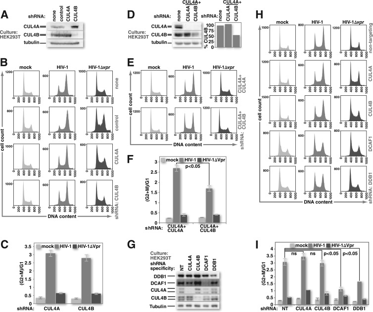
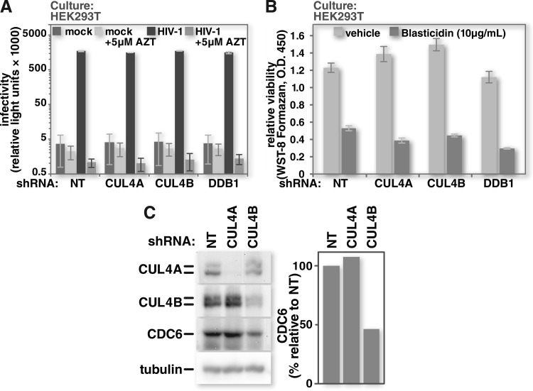
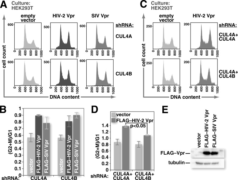
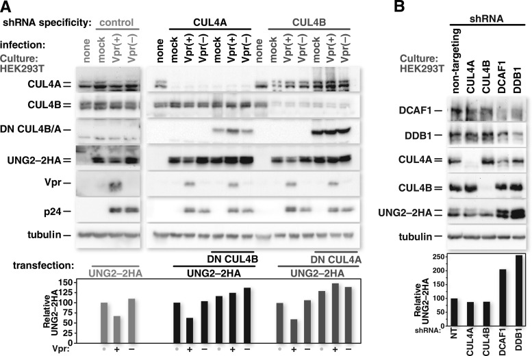
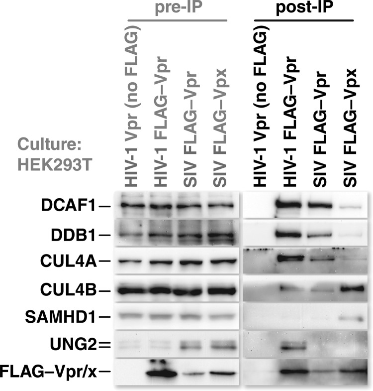
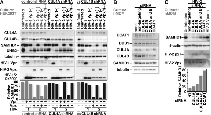
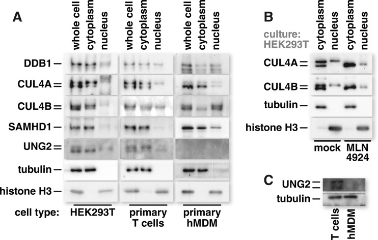
Human immunodeficiency virus (HIV) seizes control of cellular cullin-RING E3 ubiquitin ligases (CRLs) to promote viral replication. HIV-1 Vpr and HIV-2/simian immunodeficiency virus (SIV) Vpr and Vpx engage the cullin4 (CUL4)-containing ubiquitin ligase complex (CRL4) to cause polyubiquitination and proteasomal degradation of host proteins, including ones that block infection. HIV-1 Vpr engages CRL4 to trigger the degradation of uracil-N-glycosylase 2 (UNG2). Both HIV-1 Vpr and HIV-2/SIV Vpr tap CRL4 to initiate G2 cell cycle arrest. HIV-2/SIV Vpx secures CRL4 to degrade the antiviral protein SAMHD1. CRL4 includes either cullin4A (CUL4A) or cullin4B (CUL4B) among its components. Whether Vpr or Vpx relies on CUL4A, CUL4B, or both to act through CRL4 is not known. Reported structural, phenotypic, and intracellular distribution differences between the two CUL4 types led us to hypothesize that Vpr and Vpx employ these in a function-specific manner. Here we determined CUL4 requirements for HIV-1 and HIV-2/SIV Vpr-mediated G2 cell cycle arrest, HIV-1 Vpr-mediated UNG2 degradation, and HIV-2 Vpx-mediated SAMHD1 degradation. Surprisingly, CUL4A and CUL4B are exchangeable for CRL4-dependent Vpr and Vpx action, except in primary macrophages, where Vpx relies on both CUL4A and CUL4B for maximal SAMHD1 depletion. This work highlights the need to consider both CUL4 types for Vpr and Vpx functions and also shows that the intracellular distribution of CUL4A and CUL4B can vary by cell type.

Importance: The work presented here shows for the first time that HIV Vpr and Vpx do not rely exclusively on CUL4A to cause ubiquitination through the CRL4 ubiquitin ligase complex. Furthermore, our finding that intracellular CUL4 and SAMHD1 distributions can vary with cell type provides the basis for reconciling previous disparate findings regarding the site of SAMHD1 depletion. Finally, our observations with primary immune cells provide insight into the cell biology of CUL4A and CUL4B that will help differentiate the functions of these similar proteins.


Figures

FIG 1
FIG 1
HIV-1 Vpr can trigger G2 cell cycle arrest in the absence of either CUL4A or CUL4B but not both. (A) Cultures of HEK293T cells or HEK293T cells that stably express shRNA against firefly luciferase (control), CUL4A, or CUL4B were tested for CUL4-specific protein depletion by Western blotting. (B) HEK293T and HEK293T stably shRNA-expressing cell lines were mock infected or infected with HIV-1 or HIV-1 lacking Vpr at equivalent MOIs. Forty-eight hours after infection, cell nuclei were harvested, treated with RNase A and propidium iodide, and analyzed for DNA content by flow cytometry. (C) The ratio of nuclei with 4N DNA content (G2+M) to those with 2N DNA content (G1) was determined by using FlowJo software for replicates of the experiment shown in panel B. (D) HEK293T cells that stably express CUL4A-specific shRNA were depleted of CUL4B by transduction of a CUL4B-specific lentiviral expression vector. Transduction with a vector for CUL4A-specific shRNA was used as a control. Cultures under each experimental condition were tested for specific CUL4 protein depletion by Western blotting. (E) The HEK293T cultures described above for panel D were either mock infected or infected with HIV-1 or HIV-1 lacking Vpr. Nuclei from these cells were harvested 48 h after infection and analyzed for DNA content by flow cytometry, as described above for panel B. (F) The ratio of nuclei from HEK293T cells, as described above for panels D and E, with 4N DNA content to those with 2N DNA content was calculated and graphed as described above for panel C. (G) HEK293T cells with stably integrated lentiviral vectors that encode doxycycline-inducible shRNAs specific for CRL4 components were induced to express their respective shRNAs for 5 days. Transient depletion of target proteins was confirmed by Western blotting. Expression of a nontargeting (NT) shRNA served as a control. (H) HEK293T cells, as described above for panel G, were either mock infected or infected with HIV-1 or HIV-1 lacking Vpr. Forty-eight hours after infection, nuclei were isolated from these cells, and DNA content was assessed by using flow cytometry as described above for panels B and E. (I) The ratios of 4N to 2N DNA-containing nuclei, from the experiments described above for panels G and H, were calculated and graphed as described above for panels C and F (n = 3). All error bars show standard deviations. The two-tailed Student t test was used to determine statistical significance.
FIG 2
FIG 2
CRL4 depletion does not impair HIV-1 infectivity or viability of HEK293T cells. (A) HEK293T cells with stably integrated doxycycline-inducible cassettes for the expression of either a nontargeting shRNA or ones specific for CRL4 component mRNAs were induced with doxycycline. Five days after the start of induction, the cells were either mock infected or infected with VSV-G-pseudotyped HIV-1 encoding firefly luciferase in place of nef. Forty-eight hours after infection, the cells were harvested, and luciferase activity was measured by photon emission under each condition. Pretreatment of cultures with 5 μM the reverse transcription inhibitor zidovudine (AZT) served as a control to demonstrate impaired infectivity. (B) HEK293T cells were depleted of CRL4 components as described above for panel A. CRL4-depleted HEK293T cell viability was assessed by measuring water-soluble tetrazolium salt (WST-8) formazan reagent cleavage by cellular dehydrogenases. Pretreatment of cells with 10 μg/ml of the eukaryotic toxin blasticidin served as a control to demonstrate loss of viability. O.D., optical density. (C) HEK293T cells were depleted of CUL4A or CUL4B by induction of specific shRNAs. At 3 days postinduction, the cells were harvested and lysed. Cell lysates were resolved by SDS-PAGE, and Western blots were probed for CUL4A, CUL4B, CDC6, and tubulin. (n = 3 for panels A and B.) All error bars show standard deviations. The two-tailed Student t test was used to determine statistical significance.
FIG 3
FIG 3
HIV-2/SIVmac239 Vpr can trigger G2 cell cycle arrest through either CUL4A or CUL4B. (A) HEK293T cells stably expressing shRNA specific for either CUL4A or CUL4B mRNA were transfected with either an empty expression vector or one for FLAG–HIV-2 Vpr or FLAG-SIVmac239 Vpr and a small amount (7.5% of the total DNA) of an expression vector for LaminC-eGFP. Forty-eight hours after transfection, the cultures were harvested, and cell nuclei were isolated. Nuclei were treated with RNase A and stained with propidium iodide. Flow cytometry was used for gating on transfected (LaminC-eGFP-positive) cell nuclei, and DNA content was determined by measuring propidium iodide fluorescence intensity. (B) HEK293T cells that stably express shRNA specific for CUL4A were depleted of CUL4B as described in the legend of Fig. 1D. These cells were then transfected with either an empty expression vector or one for FLAG–HIV-2 Vpr. Flow cytometry was used to identify nuclei from transfected cells and to assess their DNA content as described above for panel A. (C and D) Ratios of nuclei with 4N DNA content (G2+M) to those with 2N DNA content (G1) were determined by using FlowJo software for replicates of the experiments shown in panels A (C) and B (D). (E) An empty expression vector or one for FLAG–HIV-2 Vpr or FLAG-SIVmac239 Vpr was transfected into HEK293T cells. Twenty-four hours after transfection, the cells were harvested, and expression of FLAG-tagged Vpr was confirmed by Western blot analysis with FLAG-specific antibody (n = 3). All error bars show standard deviations. The two-tailed Student t test was used to determine statistical significance.
FIG 4
FIG 4
HIV-1 Vpr-mediated UNG2 degradation and constitutive UNG2 turnover are not specifically dependent on CUL4A or CUL4B. (A) HEK293T cells that stably express a control shRNA were transfected with an expression vector for UNG2-2HA. HEK293T cells that stably express shRNA targeting CUL4A or CUL4B mRNA were transfected with an expression vector for UNG2-2HA together with either an empty expression vector or one for an HA-tagged dominant negative version of the abundant CUL4 type. Twenty-four hours after transfection, the cells were mock infected or infected with HIV-1 or HIV-1 lacking Vpr. Forty-eight hours after infection, the cells were harvested and lysed. Cell lysates were resolved by SDS-PAGE and probed for CUL4A, CUL4B, tubulin, the HA epitope, HIV-1 p24, and HIV-1 Vpr by Western blotting using the corresponding antibodies. (B) HEK293T cells stably carrying doxycycline-inducible shRNA cassettes specific for CRL4 components were induced to express their respective shRNAs for 5 days. CRL4-depleted HEK293T cells were then transfected with the UNG2-2HA expression vector and harvested 72 h after transfection. Cell lysates were resolved by SDS-PAGE, and Western blots were probed for specific CRL4 components and the HA epitope by using the corresponding antibodies. Densitometric measurements performed on the UNG2-2HA bands were normalized to the values of the respective tubulin bands. The relative intensities of the UNG2-2HA bands were graphed based on the values obtained from the densitometric analysis.
FIG 5
FIG 5
CUL4A and CUL4B are both coisolated with Vpr and Vpx. Cultures of HEK293T cells were transfected with expression vectors for untagged HIV-1 Vpr or FLAG-epitope-tagged HIV-1 Vpr, SIV Vpr, or SIV Vpx. Twenty-four hours after transfection, the cells were lysed, and insoluble debris was removed by centrifugation. Cleared lysates were incubated with FLAG-specific-antibody-coated beads to isolate the epitope-tagged viral proteins. The isolated viral proteins were subsequently eluted from the beads by competition with FLAG peptide. Proteins present in the eluates were resolved by SDS-PAGE and identified by Western blotting with specific antibodies. Preimmunoprecipitation (pre-IP) samples were derived from the lysates prior to incubation with antibody-coated beads. Untagged HIV-1 Vpr served as a control to ensure that the identified proteins were isolated through interactions with tagged viral proteins rather than nonspecific interactions with the antibody-coated beads.
FIG 6
FIG 6
Vpx can initiate SAMHD1 depletion through either CUL4A- or CUL4B-containing CRL4 complexes, but both CUL4 types are required for maximal SAMHD1 depletion in primary macrophages. (A) HEK293T cells that stably express a control shRNA or shRNAs specific for CUL4A or CUL4B mRNAs were either mock infected or infected at equivalent MOIs with HIV-1, HIV-1 lacking Vpr, HIV-2, or HIV-2 lacking Vpr, Vpx, or both. Twenty-four hours after infection, the cells were collected and lysed. Proteins from the whole-cell lysates were resolved by SDS-PAGE and identified by Western blotting using protein-specific antibodies. (B) Cultures of primary human monocyte-derived macrophages (hMDMs) were either mock transfected or transfected with nontargeting siRNA or siRNA specific for the mRNA of the CRL4 complex components indicated. Protein depletion was confirmed by Western blotting. (C, top) Forty-eight hours after siRNA transfection, the cells were either mock infected or infected at a high MOI (MOI = 10) with HIV-2 or HIV-2 lacking Vpx. Less than 24 h after infection, the cells were harvested and lysed. Proteins from whole-cell lysates were resolved by SDS-PAGE and identified by Western blotting with protein-specific antibodies. (Bottom) Densitometric measurements performed on the SAMHD1 bands were normalized to the values of the respective β-actin bands. The relative intensities of the SAMHD1 bands were graphed based on the values obtained from the densitometric analysis. Data shown are representative of a trend observed for samples from 3 different donors.
FIG 7
FIG 7
Intracellular CUL4A and CUL4B distributions can vary by cell type. (A) Cultures of HEK293T cells, primary T cells, and primary hMDMs were separated into cytosolic and nuclear fractions by cell membrane lysis and centrifugation of nuclei through a sucrose cushion. Proteins present in the cytosolic or nuclear fraction were resolved by SDS-PAGE and identified by Western blotting using antibodies with the indicated specificities. Tubulin and histone signals were used to confirm the purity of cytosolic and nuclear fractions, respectively, and sample loading was normalized to the signal from whole-cell lysates to ensure an equal representation of cytoplasmic and nuclear lysates. (B) HEK293T cells were pretreated with 1 μM the neddylation inhibitor MLN4924 for 24 h before the cells were harvested and cytosolic and nuclear fractions were isolated. Proteins from cellular fractions were resolved by SDS-PAGE and identified by Western blotting probing for CUL4A, CUL4B, tubulin, and histone. (C) Proteins from the whole-cell lysates of primary T cells and primary hMDMs were resolved by SDS-PAGE, and the levels of tubulin and endogenous UNG2 were assessed by Western blotting. Data shown are representative of a trend observed for 2 different donors.

Similar articles

Cited by

References

    1. Yu X, Yu Y, Liu B, Luo K, Kong W, Mao P, Yu XF. 2003. Induction of APOBEC3G ubiquitination and degradation by an HIV-1 Vif-Cul5-SCF complex. Science 302:1056–1060. 10.1126/science.1089591 - DOI - PubMed
    1. Stopak K, de Noronha C, Yonemoto W, Greene WC. 2003. HIV-1 Vif blocks the antiviral activity of APOBEC3G by impairing both its translation and intracellular stability. Mol. Cell 12:591–601. 10.1016/S1097-2765(03)00353-8 - DOI - PubMed
    1. Sheehy AM, Gaddis NC, Malim MH. 2003. The antiretroviral enzyme APOBEC3G is degraded by the proteasome in response to HIV-1 Vif. Nat. Med. 9:1404–1407. 10.1038/nm945 - DOI - PubMed
    1. Marin M, Rose KM, Kozak SL, Kabat D. 2003. HIV-1 Vif protein binds the editing enzyme APOBEC3G and induces its degradation. Nat. Med. 9:1398–1403. 10.1038/nm946 - DOI - PubMed
    1. Conticello SG, Harris RS, Neuberger MS. 2003. The Vif protein of HIV triggers degradation of the human antiretroviral DNA deaminase APOBEC3G. Curr. Biol. 13:2009–2013. 10.1016/j.cub.2003.10.034 - DOI - PubMed

Publication types

MeSH terms

Substances