. 2014 Jul;88(13):7130-44.
doi: 10.1128/JVI.00420-14. Epub 2014 Apr 9.

Conserved neutralizing epitope at globular head of hemagglutinin in H3N2 influenza viruses

Affiliations

Conserved neutralizing epitope at globular head of hemagglutinin in H3N2 influenza viruses

Yoshitaka Iba et al. J Virol. 2014 Jul.

Abstract

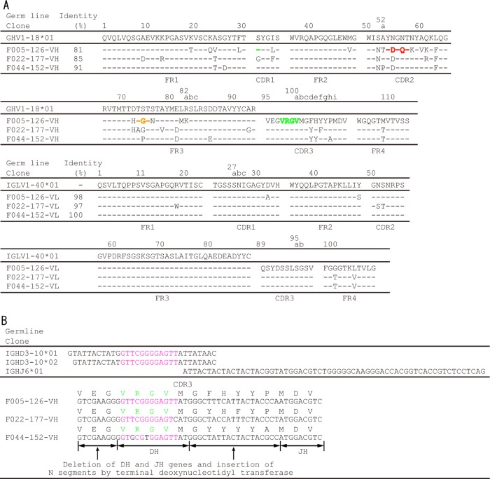
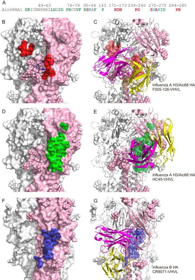
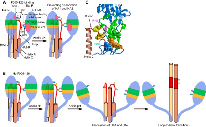
Neutralizing antibodies that target the hemagglutinin of influenza virus either inhibit binding of hemagglutinin to cellular receptors or prevent the low-pH-induced conformational change in hemagglutinin required for membrane fusion. In general, the former type of antibody binds to the globular head formed by HA1 and has narrow strain specificity, while the latter type binds to the stem mainly formed by HA2 and has broad strain specificity. In the present study, we analyzed the epitope and function of a broadly neutralizing human antibody against H3N2 viruses, F005-126. The crystal structure of F005-126 Fab in complex with hemagglutinin revealed that the antibody binds to the globular head, spans a cleft formed by two hemagglutinin monomers in a hemagglutinin trimer, and cross-links them. It recognizes two peptide portions (sites L and R) and a glycan linked to asparagine at residue 285 using three complementarity-determining regions and framework 3 in the heavy chain. Binding of the antibody to sites L (residues 171 to 173, 239, and 240) and R (residues 91, 92, 270 to 273, 284, and 285) is mediated mainly by van der Waals contacts with the main chains of the peptides in these sites and secondarily by hydrogen bonds with a few side chains of conserved sequences in HA1. Furthermore, the glycan recognized by F005-126 is conserved among H3N2 viruses. F005-126 has the ability to prevent low-pH-induced conformational changes in hemagglutinin. The newly identified conserved epitope, including the glycan, should be immunogenic in humans and may induce production of broadly neutralizing antibodies against H3 viruses.

Importance: Antibodies play an important role in protection against influenza virus, and hemagglutinin is the major target for virus neutralizing antibodies. It has long been believed that all effective neutralizing antibodies bind to the surrounding regions of the sialic acid-binding pocket and inhibit the binding of hemagglutinin to the cellular receptor. Since mutations are readily introduced into such epitopes, this type of antibody shows narrow strain specificity. Recently, however, broadly neutralizing antibodies have been isolated. Most of these bind either to conserved sites in the stem region or to the sialic acid-binding pocket itself. In the present study, we identified a new neutralizing epitope in the head region recognized by a broadly neutralizing human antibody against H3N2. This epitope may be useful for design of vaccines.


Figures

FIG 1
FIG 1
Amino acid sequences of F005-126 VH and VL. (A) Amino acid numbers are according to the Kabat numbering system. Framework regions FR1, FR2, FR3, and FR4 and complementary determining regions CDR1, CDR2, and CDR3 are shown. Comparison of amino acid sequences of VHs and VLs to germ line sequences was performed according to IgBLAST (http://www.ncbi.nlm.nih.gov/igblast/). Amino acid sequence identities (%) of MAbs with germ line VH or VL are shown. From X-ray analysis, amino acid sequences indicated in orange, green, and red were bound to site L, site R, and a glycan at HA-R:Asn285, respectively. (B) Predicted deletion of DH and JH genes and insertion of N segments are shown. The nucleotide sequence GTTCGGGGAGTT, encoding the 4-amino-acid sequence V98–R99-G100–V100a in F005-126–HCDR3, is indicated in pink.
FIG 2
FIG 2
Neutralizing and binding activities of F005-126. (A) Virus-neutralizing activity against 12 H3N2 strains was measured by focus reduction assay (31). (B) Binding activity of Abs to HA artificially expressed on cells was examined by flow cytometry (FCM). Blue line, HA of Fuk85 (H3N2); red line, HA of NC99 (H1N1); gray filled, mock transfection. F045-092 binds to both H3-type and H1-type HAs (24). F49 binds to an epitope on HA2 commonly present in H3 HA (33). C179 binds to the stem region of HA of group 1 virus. Anti-V5 Ab binds to the V5 tag located at the membrane-proximal end of HA. (C) Binding activity of Abs to HA (blue) and HA1 domain (red) of Fuk85 was analyzed by FCM. F019-102 binds to site E on HA1 (35). (D) Binding activity of Abs to HA (blue) and truncated HAs, HA39-319 (red) and HA44-309 (orange) of Fuk85 was analyzed by FCM. Anti-myc Ab binds to the myc tag located at the membrane-proximal end of HA.
FIG 3
FIG 3
Competition for binding to HA between F005-126 and four MAbs that bind to sites C1/C2, E, and B1 on HA1. Competitive ELISA for Yam77 virus particles was performed by using Fab-PP as primary Ab for detection of binding activity without (−) (gray) or with (+) (red or blue) a large excess of competitor Fab-cp3 (for example, F041-342cp3). Fab-PP is Fab fused with IgG-binding domain of protein A, and Fab-cp3 is Fab fused with coat protein 3 (cp3) of filamentous phage. Fab-PP was detected by peroxidase-conjugated rabbit Ab. The antigenic sites recognized by the four MAbs—F041-342, F041-360, F019-102, and F037-115—are indicated in parentheses above the graphs.
FIG 4
FIG 4
Neutralization and binding activity of F005-126 against mutant Aic68/N285Y. (A) Neutralization activity of F005-126 against an escape mutant, Aic68N285Y, was weaker than that against the wild-type Aic68 strain. (B) FCM analyses of the cells expressing Aic68HA/wild-type and a mutant Aic68HA/N285Y were performed. FCM signals for mock transfection (gray filled), wild-type HA-expressing cells (blue), and mutant HA-expressing cells (red) are shown. F045-092, F003-137, and F035-015 recognize the HA head region (24, 34). Binding activity of F005-126 to Aic68HA/N285Y was weaker than that to wild-type HA.
FIG 5
FIG 5
Amino acids around site C, site E, and Asn285 in HA1 on the 3D structure of H3 HA. Residues 50 to 57 and residues 275 to 279 in antigenic site C are indicated in yellow. Residues 62 to 83 in antigenic site E are indicated in violet. Residues 39 to 43, 310 to 316, and 285 in HA1 are indicated in green, cyan, and red, respectively. Residues 9 to 38 and residues 317 to 329 in HA1 are indicated in orange. Residues 44 to 309 are indicated in blue. The HA2 domain is gray. A glycan at Asn285 is white.
FIG 6
FIG 6
Crystal structure of F005-126 Fab in complex with Aic68/HA. (A) One HA trimer forms a complex with three F005-126 Fabs as indicated. In an asymmetric unit (AU), the crystal contains 12 F005-126 Fabs and four Aic68/HA trimers (twelve Aic68/HA monomers). Three Fabs bind to the HA trimer in the same manner. CH1/CL domains in 12 Fabs curve in the different patterns. (B to E) The figures show 2Fo-Fc electron density maps of the helix of residues 79 to 94 in HA2 (B), FR3 (C), CDR3 (D), and CDR2 (E). Fo means observed structure factor and Fc means calculated structure factor. The electron density is contoured at 1.5 σ and shown by a 12-fold noncrystallographically averaged map.
FIG 7
FIG 7
Crystal structure of F005-126 Fab in complex with Aic68/HA. (A and B) Top and side views, respectively, of the crystal structure. Trimeric HAs are depicted in a surface representation (A) and by ribbons (B). HA-L, HA1-R, HA2-R, and an HA in an HA trimer are indicated in light yellow, lavender, blue, and white, respectively. Sites L and R are shown in red. F005-126 Fabs are shown as ribbons. One Fab is colored. The H and L chains are green and magenta, respectively. Glycans are depicted as spheres. (C) Closeup view of sites L and R. A glycan at Asn285 in HA1-R is depicted as lavender spheres. CDR1, CDR2, CDR3, and FR3 of the H chain are depicted as wire frames in gray, magenta, green, and orange, respectively. Sites L and R are indicated in red.
FIG 8
FIG 8
Crystal structure of F005-126 Fab in complex with Aic68/HA. (A and B) Epitopes on HA recognized by F005-126 are shown. In panel B, putative hydrogen bonds are depicted as broken lines.
FIG 9
FIG 9
Interactions between F005-126 Fab and Aic68/HA in the crystal. (A) Interactions between site L and FR3 of the H chain are shown. (B) Interactions among site R, the glycan at HA1-R:Asn285, HCDR2 and HCDR3 are shown. N-acetyl-d-glucosamine, α-d-mannose, and β-d-mannose are abbreviated as NAG, MAN, and BMA, respectively. Putative hydrogen bonds are depicted as broken lines. (C) The interactions in Fig. 7B are schematically depicted.
FIG 10
FIG 10
Inhibition of the low-pH-induced conformational changes by binding of F005-126 to HA1s. (A) HA was digested by trypsin after incubation at low pH (lane 3), but F005-126 prevented the low-pH-induced conformational change and thus rendered HA resistant to trypsin (lane 7). (B) HA1-L, HA2-L, HA1-R, and HA2-R are depicted in green, light brown, blue, and dark brown, respectively, except for the B loop. The B loops in HA2-L and HA2-R are depicted in red. The following residues in sites L and R are shown as red spheres: residues 171 to 173, 239, and 240 in HA1-L and residues 91, 92, 270 to 273, 284, and 285 in HA1-R. Glycans are depicted as sticks. The following amino acid residues are shown as spheres: K238 in HA1-L (cyan); K27, R109, R269, and K310 in HA1-R (cyan); K62 in HA2-L (cyan); and E67, E72, D86, D90, and E97 in HA2-R (yellow). [HA1-R:R109]-[HA2-R:E67]-[HA1-R:R269] in H3N2 make salt bridges (57). The crystal structure of H3N2 HA also shows the other putative salt bridges made by the following amino acids: [HA1-L:K238]-[HA2-R:E72], [HA2-L:K62]-[HA2-R:D86D90]-[HA1-R:K310], and [HA1-R:K27]-[HA2-R:E97].
FIG 11
FIG 11
Antigenic sites in HA trimers of H1N1/SC1918 and H3N2/Aic68. HA1s and HA2s in HA trimers are white and brown, respectively. (Left) Antigenic sites Sa (blue), Sb (cyan), Ca1 (orange), Ca2 (green), and Cb (violet) in two of three HAs from H1N1/SC1918 are indicated in colors, and a region corresponding to site R is red. (Right) Antigenic sites A (blue), B (cyan), C (yellow), D in the left side of HA (orange), D in the right side of HA (green), and E (violet) in two of three HAs from H3N2/Aic68 are colored as indicated, and sites L and R are red.
FIG 12
FIG 12
Comparison of epitopes recognized by F005-126, HC45, and CR8071. (A) The amino acid sequences of Aic68 HA recognized by F005-126 (red), HC45 (green), and both Abs (cyan) are colored as indicated. (B to G) An HA protomer in each HA trimer is light pink. The epitopes in HA of Aic68 recognized by F005-126 (B and C) and HC45 (PDB ID 1QFU) (D and E) are indicated in red and green, respectively. The amino acid numbers in HA of influenza virus A Aic68 strain are shown in panels B and D. The epitope in HA of influenza virus B recognized by CR8071 (PDB ID 4FQK) is blue (F and G). The amino acid numbers in HA of influenza virus B, which do not correspond to that in HA of influenza virus A H3, are shown. The VH and VL domains are magenta and yellow, respectively (C, E, and G).
FIG 13
FIG 13
Illustrations on a proposed mechanism for preventing dissociation of HA1 and HA2 by F005-126. (A and B) HA1, helix A, B loop and helix C in two HA protomers in an HA trimer are illustrated. A vestigial esterase (VE) subdomain is green (residues 52 to 116) and orange (residues 266 to 275), and other regions in HA1 are blue. Arg109 and Arg269 are violet and cyan, respectively. Glu67 and Glu72 in B loop are yellow. Sites L and R are indicated in magenta. Under the low-pH condition, initially, part of the salt bridges are broken, and finally, the salt bridges are completely broken. When F005-126 binds to HA, dissociation does not occur at low pH, even if the salt bridges are broken (A). Dissociation of HA1 and HA2 occurs at low pH without F005-126 (B). (C) Arg109 and Arg269 are depicted as sticks in violet and cyan, respectively. Glu67 and Glu72 in B loop are depicted as surface representation in yellow. Residues 91, 92, 270 to 273, 284, and 285 in site R are depicted as surface representations.

References

    1. Skehel JJ, Wiley DC. 2000. Receptor binding and membrane fusion in virus entry: the influenza hemagglutinin. Annu. Rev. Biochem. 69:531–569. 10.1146/annurev.biochem.69.1.531 - DOI - PubMed
    1. Wiley DC, Wilson IA, Skehel JJ. 1981. Structural identification of the antibody-binding sites of Hong Kong influenza haemagglutinin and their involvement in antigenic variation. Nature 289:373–378. 10.1038/289373a0 - DOI - PubMed
    1. Underwood PA. 1982. Mapping of antigenic changes in the haemagglutinin of Hong Kong influenza (H3N2) strains using a large panel of monoclonal antibodies. J. Gen. Virol. 62:153–169. 10.1099/0022-1317-62-1-153 - DOI - PubMed
    1. Caton AJ, Brownlee GG, Yewdell JW, Gerhard W. 1982. The antigenic structure of the influenza virus A/PR/8/34 hemagglutinin (H1 subtype). Cell 31:417–427. 10.1016/0092-8674(82)90135-0 - DOI - PubMed
    1. Salzberg S. 2008. The contents of the syringe. Nature 454:160–161. 10.1038/454160a - DOI - PMC - PubMed

Publication types

MeSH terms

Substances