

Postnatal acquisition of primary rhesus cytomegalovirus infection is associated with prolonged virus shedding and impaired CD4+ T lymphocyte function
- PMID: 24719473
- PMCID: PMC4215082
- DOI: 10.1093/infdis/jiu215
Postnatal acquisition of primary rhesus cytomegalovirus infection is associated with prolonged virus shedding and impaired CD4+ T lymphocyte function
Abstract
Background: Although virus-specific CD4(+) T lymphocytes emerge rapidly during primary cytomegalovirus (CMV) infection in humans, they exhibit a state of prolonged functional exhaustion of unknown etiology. To investigate the suitability of rhesus macaques as a model of primary human CMV infection, we examined the virologic and immunologic features of naturally acquired primary CMV infection in rhesus macaques.
Methods: CMV-specific CD4(+) T lymphocytes and CMV load in blood, saliva, and urine were evaluated in a cohort of simian immunodeficiency virus (SIV)-negative rhesus macaques stratified by age into infant, juvenile, and adult groups.
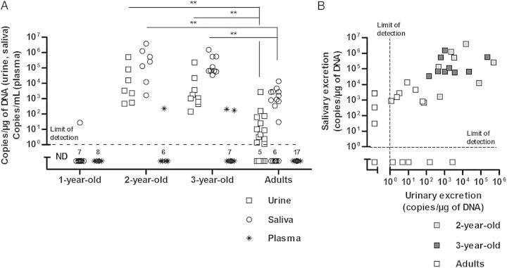
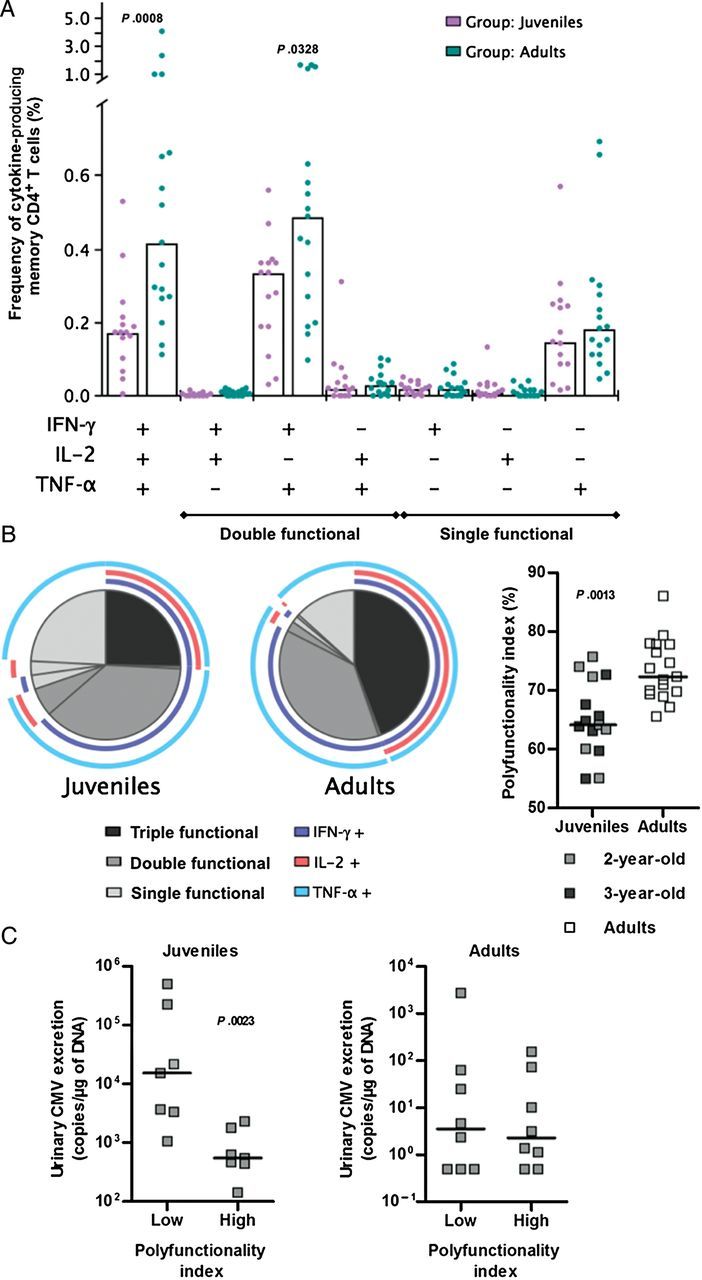
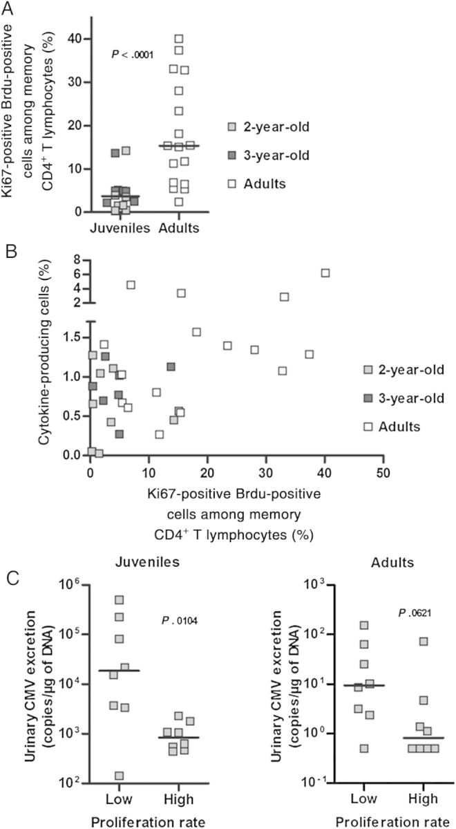
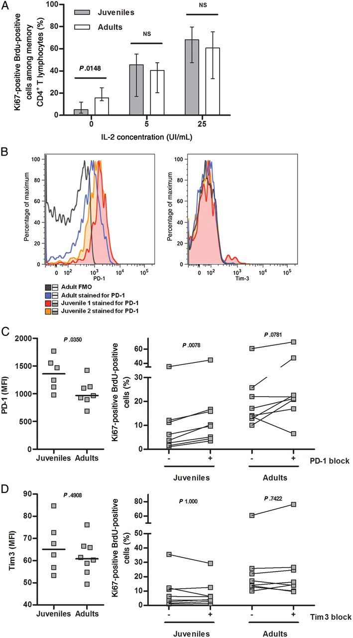
Results: CMV infection was detected in juvenile and adult monkeys but not in infant monkeys. CMV loads and shedding frequency in urine and saliva were significantly higher in the 2-3-year old juvenile monkeys, compared with the adult monkeys. The increased CMV load in juvenile monkeys was associated with lower polyfunctionality, impaired proliferation, and increased expression of the inhibitory receptor PD-1 in CMV-specific CD4(+) T lymphocytes. The proliferative defect was partially reversible by exogenous PD-1 blockade or addition of interleukin 2.
Conclusions: Postnatal acquisition of primary CMV infection in rhesus macaques results in prolonged virus excretion and impaired CMV-specific CD4(+) T-lymphocyte function, findings that recapitulate key features of primary CMV infection in humans.
Keywords: CD4+ T lymphocyte; IFN-γ; IL-2; PD-1; TNF-α; cytomegalovirus; functional exhaustion; primary cytomegalovirus infection; rhesus macaque; viral excretion.
© The Author 2014. Published by Oxford University Press on behalf of the Infectious Diseases Society of America. All rights reserved. For Permissions, please e-mail: journals.permissions@oup.com.
Figures

Similar articles
-
Decreased frequency of cytomegalovirus (CMV)-specific CD4+ T lymphocytes in simian immunodeficiency virus-infected rhesus macaques: inverse relationship with CMV viremia.J Virol. 2002 Apr;76(8):3646-58. doi: 10.1128/jvi.76.8.3646-3658.2002. J Virol. 2002. PMID: 11907204 Free PMC article.
-
Immunohistochemical studies of productive rhesus cytomegalovirus infection in rhesus monkeys (Macaca mulatta) infected with simian immunodeficiency virus.Vet Pathol. 1999 Jan;36(1):51-6. doi: 10.1354/vp.36-1-51. Vet Pathol. 1999. PMID: 9921756
-
Persistent and selective deficiency of CD4+ T cell immunity to cytomegalovirus in immunocompetent young children.J Immunol. 2004 Mar 1;172(5):3260-7. doi: 10.4049/jimmunol.172.5.3260. J Immunol. 2004. PMID: 14978134
-
Direct relationship between suppression of virus-specific immunity and emergence of cytomegalovirus disease in simian AIDS.J Virol. 2003 May;77(10):5749-58. doi: 10.1128/jvi.77.10.5749-5758.2003. J Virol. 2003. PMID: 12719568 Free PMC article.
-
Urinary cytomegalovirus excretion: The unresolved issues.Ann Pharm Fr. 2024 Sep;82(5):755-761. doi: 10.1016/j.pharma.2024.03.004. Epub 2024 Mar 16. Ann Pharm Fr. 2024. PMID: 38492661 Review.
Cited by
-
Human cytomegalovirus (HCMV) long-term shedding and HCMV-specific immune response in pregnant women with primary HCMV infection.Med Microbiol Immunol. 2022 Dec;211(5-6):249-260. doi: 10.1007/s00430-022-00747-4. Epub 2022 Aug 12. Med Microbiol Immunol. 2022. PMID: 35960328
-
The Cellular Localization of Human Cytomegalovirus Glycoprotein Expression Greatly Influences the Frequency and Functional Phenotype of Specific CD4+ T Cell Responses.J Immunol. 2015 Oct 15;195(8):3803-15. doi: 10.4049/jimmunol.1500696. Epub 2015 Sep 11. J Immunol. 2015. PMID: 26363059 Free PMC article. Clinical Trial.
-
Characterization of Baboon Cytomegalovirus Infection in Healthy Adult Baboons (Papio anubis).Comp Med. 2019 Feb 1;69(1):55-62. doi: 10.30802/AALAS-CM-18-000050. Epub 2019 Jan 31. Comp Med. 2019. PMID: 30704552 Free PMC article.
-
Rhesus monkeys for a nonhuman primate model of cytomegalovirus infections.Curr Opin Virol. 2017 Aug;25:126-133. doi: 10.1016/j.coviro.2017.08.005. Epub 2017 Sep 6. Curr Opin Virol. 2017. PMID: 28888133 Free PMC article. Review.
-
Protective effect of pre-existing natural immunity in a nonhuman primate reinfection model of congenital cytomegalovirus infection.PLoS Pathog. 2023 Oct 5;19(10):e1011646. doi: 10.1371/journal.ppat.1011646. eCollection 2023 Oct. PLoS Pathog. 2023. PMID: 37796819 Free PMC article.
References
-
- Gamadia LE, Remmerswaal EB, Weel JF, Bemelman F, van Lier RA, Ten Berge IJ. Primary immune responses to human CMV: a critical role for IFN-gamma-producing CD4+ T cells in protection against CMV disease. Blood. 2003;101:2686–92. - PubMed
-
- Pourgheysari B, Piper KP, McLarnon A, et al. Early reconstitution of effector memory CD4+ CMV-specific T cells protects against CMV reactivation following allogeneic SCT. Bone Marrow Transplant. 2009;43:853–61. - PubMed
-
- Appay V, Zaunders JJ, Papagno L, et al. Characterization of CD4(+) CTLs ex vivo. J Immunol. 2002;168:5954–8. - PubMed
Publication types
MeSH terms
Grants and funding
LinkOut - more resources
Full Text Sources
Other Literature Sources
Medical
Research Materials
