. 2014:2014:208103.
doi: 10.1155/2014/208103. Epub 2014 Feb 27.

Interleukins affect equine endometrial cell function: modulatory action of ovarian steroids

Affiliations

Interleukins affect equine endometrial cell function: modulatory action of ovarian steroids

Anna Z Szóstek et al. Mediators Inflamm. 2014.

Abstract

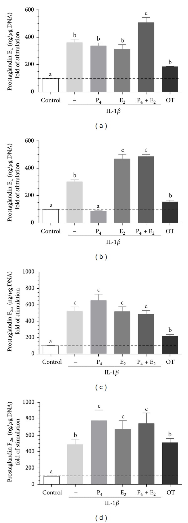
The aim of the present study was to investigate the interaction between ovarian steroids, interleukins and prostaglandins (PG) in equine epithelial and stromal cells in vitro. In Experiment 1, cells were exposed to IL-1α (10 ng/mL), IL-1β (10 ng/mL) or IL-6 (10 ng/mL) for 24 h and cell proliferation was determined using MTT. In Experiment 2, cells were exposed to progesterone (P4; 10(-7) M); 17-β estradiol (E2; 10(-9) M) or P4+E2 for 24 h and later medium was replaced with a fresh one treated with IL-1α, IL-1β or IL-6 (10 ng/mL, each) for 24 h. The oxytocin (OT; 10(-7) M) was used as a positive control. In Experiment 3, cells were exposed to P4 (10(-7) M), E2 (10(-9) M) or P4+E2 for 24 h and the IL receptor mRNAs transcription was determined using Real-time PCR. Prostaglandins concentration was determined using the direct enzyme immunoassay (EIA) method. Our findings reveal a functional linking between ovarian steroids and IL-stimulated PG secretion by equine endometrial cells. This interaction could be one of the mechanisms responsible for endometrial local orchestrating events during the estrous cycle and early pregnancy.


Figures

Figure 1
Figure 1
Representative morphology of cultured equine endometrial: (a) epithelial cells and (b) stromal cells. (c) Epithelial cells identification by immunofluorescent staining for cytokeratin; (d) stromal cells identification by immunofluorescent staining for vimentin. The scale bar = 50 μm (magnification: ×40).
Figure 2
Figure 2
The effect of IL-1α (10 ng/mL), IL-1β (10 ng/mL), or IL-6 (10 ng/mL) on proliferation of (a) epithelial cells (mean ± SEM; n = 5) and (b) stromal cells (mean ± SEM; n = 5) after 24 h incubation. All values are expressed as n-fold change from control. Asterisks indicate significant differences (*P < 0.05; **P < 0.01) from the respective control, as determined by parametric one-way ANOVA followed by Newmann-Keuls comparison test.
Figure 3
Figure 3
The effect of ovarian steroids on IL-1α stimulated PGE2 and PGF2α production by epithelial ((a), (c)) and stromal ((b), (d)) cells. The cells were treated with P4 (10−7 M), E2 (10−9 M), or P4 + E2 (10−7/10−9 M) for 24 h. Then, the medium was replaced with a fresh medium and the epithelial cells were stimulated with IL-1α (10 ng/mL) for 24 h. Oxytocin (OT; 10−7 M) was used as a positive control. All values are expressed as n-fold change from control. letters “a,” “b,” and “c” indicate significant differences (P < 0.05) between groups, as determined by parametric one-way ANOVA followed by Newmann-Keuls comparison test.
Figure 4
Figure 4
The effect of ovarian steroids on IL-1β stimulated PGE2 and PGF2α production by epithelial ((a), (c)) and stromal ((b), (d)) cells. The cells were treated with P4 (10−7 M), E2 (10−9 M), or P4 + E2 (10−7/10−9 M) for 24 h. Then, the medium was replaced with a fresh medium and the epithelial cells were stimulated with IL-1β (10 ng/mL) for 24 h. Oxytocin (OT; 10−7 M) was used as a positive control. All values are expressed as n-fold change from control. letters “a,” “b,” “c” indicate significant differences (P < 0.05) between groups, as determined by parametric one-way ANOVA followed by Newmann-Keuls comparison test.
Figure 5
Figure 5
The effect of ovarian steroids on IL-6 stimulated PGE2 and PGF2α production by epithelial ((a), (c)) and stromal ((b), (d)) cells. The cells were treated with P4 (10−7 M), E2 (10−9 M), or P4 + E2 (10−7/10−9 M) for 24 h. Then, the medium was replaced with a fresh medium and the epithelial cells were stimulated with IL-6 (10 ng/mL) for 24 h. Oxytocin (OT; 10−7 M) was used as a positive control. All values are expressed as n-fold change from control. letters “a,” “b,” and “c” indicate significant differences (P < 0.05) between groups, as determined by parametric one-way ANOVA followed by Newmann-Keuls comparison test.
Figure 6
Figure 6
The effect of P4 (10−7 M), E2 (10−9 M), and P4 + E2 (10−7/10−9 M) on IL-1RI ((a), (b)), IL-1RII ((c), (d)), and IL-6Rβ ((e), (f)) mRNA transcription in epithelial cells (n = 5) and stromal cells (n = 5) after 24 h incubation. Results are normalized against ACTB. Data are presented as fold induction relative to control. Asterisks indicate significant differences (*P < 0.05; **P < 0.01; ***P < 0.001) from the respective control, as determined by nonparametric one-way ANOVA Kruskala-Wallisa followed by Dunns'a test.
Figure 7
Figure 7
The effect of P4 (10−7 M), E2 (10−9 M), and P4 + E2 (10−7/10−9 M) on IL-1RI:IL-1RII mRNA transcription ratio in epithelial cells ((a); n = 5) and stromal cells ((b); n = 5) after 24 h incubation. Results are normalized against ACTB. Asterisks indicate significant differences (*P < 0.05; **P < 0.01) from the respective control, as determined by nonparametric one-way ANOVA Kruskala-Wallisa followed by Dunns'a test.

References

    1. Vernon MW, Zavy MT, Asquith RL, Sharp DC. Prostaglandin F2(α) in the equine endometrium: steroid modulation and production capacities during the estrous cycle and early pregnancy. Biology of Reproduction. 1981;25(3):581–589. - PubMed
    1. Zavy MT, Vernon MW, Asquith RL. Effect of exogenous gonadal steroids and pregnancy on uterine luminal prostaglandin F in mares. Prostaglandins. 1984;27(2):311–320. - PubMed
    1. Galvão A, Valente L, Skarzynski DJ, et al. Effect of cytokines and ovarian steroids on equine endometrial function: an in vitro study. Reproduction, Fertility and Development. 2013;25(7):985–997. - PubMed
    1. Szóstek AZ, Galvão AM, Ferreira-Dias GM, Skarzynski DJ. Ovarian steroids affect prostaglandin production in equine endometrial cells in vitro . Journal of Endocrinology. 2014;220(3):267–276. - PubMed
    1. Skarzynski DJ, Bah MM, Deptula KM, et al. Roles of tumor necrosis factor-α of the estrous cycle in cattle: an in vivo study. Biology of Reproduction. 2003;69(6):1907–1913. - PubMed

Publication types

LinkOut - more resources