

Adenovirus-mediated prothymosin α gene transfer inhibits the development of atherosclerosis in ApoE-deficient mice
- PMID: 24719553
- PMCID: PMC3979988
- DOI: 10.7150/ijbs.8634
Adenovirus-mediated prothymosin α gene transfer inhibits the development of atherosclerosis in ApoE-deficient mice
Abstract
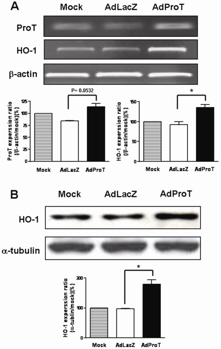
Prothymosin α (ProT) is involved in regulating expression of the oxidative stress-protective genes and it also exerts immunomodulatory activities. In this study, we investigated the therapeutic effects of ProT gene transfer on atherosclerosis in endothelial cells and in ApoE-deficient mice. Adenoviruses encoding mouse ProT (AdProT) were used for the management of atherosclerosis. In vitro, the effects of ProT on antioxidant gene expressions and the protection effect against oxidant-mediated injury in endothelial cells were examined. In vivo, AdProT were administered intraventricularly into the heart of ApoE(-/-) mice. Histopathological and immunohistochemical assessments of the aortic tissues were performed. Expressions of HO-1 and antioxidant genes in the aortic tissues were also determined. Our results demonstrated that ProT gene transfer increased antioxidant gene expressions, eNOS expression and NO release, as well as reduced the reactive oxygen species production in endothelial cells. Intraventricular administration of AdProT reduced the lesion formation, increased expressions of HO-1 and SOD genes, and reduced infiltrating macrophages in the aorta of ApoE(-/-) mice. This study suggests that ProT gene transfer may have the therapeutic potential for the management of atherosclerosis via inducing antioxidant gene expressions, eNOS expression and NO release, reducing ROS production and macrophage infiltration in endothelium.
Keywords: antioxidant gene expressions; atherosclerosis; gene transfer; prothymosin α..
Conflict of interest statement
Competing Interests: The authors have declared that no competing interest exists.
Figures


Similar articles
-
Specific dietary polyphenols attenuate atherosclerosis in apolipoprotein E-knockout mice by alleviating inflammation and endothelial dysfunction.Arterioscler Thromb Vasc Biol. 2010 Apr;30(4):749-57. doi: 10.1161/ATVBAHA.109.199687. Epub 2010 Jan 21. Arterioscler Thromb Vasc Biol. 2010. PMID: 20093625
-
BRCA1 is a novel target to improve endothelial dysfunction and retard atherosclerosis.J Thorac Cardiovasc Surg. 2013 Oct;146(4):949-960.e4. doi: 10.1016/j.jtcvs.2012.12.064. Epub 2013 Feb 14. J Thorac Cardiovasc Surg. 2013. PMID: 23415688
-
IGF-1 reduces inflammatory responses, suppresses oxidative stress, and decreases atherosclerosis progression in ApoE-deficient mice.Arterioscler Thromb Vasc Biol. 2007 Dec;27(12):2684-90. doi: 10.1161/ATVBAHA.107.156257. Epub 2007 Oct 4. Arterioscler Thromb Vasc Biol. 2007. PMID: 17916769
-
β-Elemene attenuates atherosclerosis in apolipoprotein E-deficient mice via restoring NO levels and alleviating oxidative stress.Biomed Pharmacother. 2017 Nov;95:1789-1798. doi: 10.1016/j.biopha.2017.08.092. Epub 2017 Oct 6. Biomed Pharmacother. 2017. PMID: 28962084
-
Irisin protects against endothelial injury and ameliorates atherosclerosis in apolipoprotein E-Null diabetic mice.Atherosclerosis. 2015 Dec;243(2):438-48. doi: 10.1016/j.atherosclerosis.2015.10.020. Epub 2015 Oct 19. Atherosclerosis. 2015. PMID: 26520898
Cited by
-
Oxidative Stress in Cardiovascular Diseases: Involvement of Nrf2 Antioxidant Redox Signaling in Macrophage Foam Cells Formation.Int J Mol Sci. 2017 Nov 5;18(11):2336. doi: 10.3390/ijms18112336. Int J Mol Sci. 2017. PMID: 29113088 Free PMC article. Review.
-
MiRNA Let-7i-5p-Contained Small Extracellular Vesicles from Macrophages Induce Nucleus Pulposus Cell Senescence via Targeting LIN28A.Int J Nanomedicine. 2025 Feb 18;20:2163-2179. doi: 10.2147/IJN.S482646. eCollection 2025. Int J Nanomedicine. 2025. PMID: 39990291 Free PMC article.
-
Transient Receptor Potential Ankyrin 1 Channel Involved in Atherosclerosis and Macrophage-Foam Cell Formation.Int J Biol Sci. 2016 May 25;12(7):812-23. doi: 10.7150/ijbs.15229. eCollection 2016. Int J Biol Sci. 2016. PMID: 27313495 Free PMC article.
-
Prothymosin α Gene Transfer Modulates Myocardial Remodeling after Ischemia-Reperfusion Injury.Acta Cardiol Sin. 2022 Mar;38(2):187-200. doi: 10.6515/ACS.202203_38(2).20211115A. Acta Cardiol Sin. 2022. PMID: 35273440 Free PMC article.
-
Prothymosin α accelerates dengue virus-induced thrombocytopenia.iScience. 2023 Dec 2;27(1):108422. doi: 10.1016/j.isci.2023.108422. eCollection 2024 Jan 19. iScience. 2023. PMID: 38213625 Free PMC article.
References
-
- Stocker R, Keaney JF Jr. Role of oxidative modifications in atherosclerosis. Physiol Rev. 2004;84:1381–478. - PubMed
-
- Madamanchi NR, Vendrov A, Runge MS. Oxidative stress and vascular disease. Arterioscler Thromb Vasc Biol. 2005;25:29–38. - PubMed
-
- Yokoyama M. Oxidant stress and atherosclerosis. Curr Opin Pharmacol. 2004;4:110–5. - PubMed
-
- Faraci FM, Didion SP. Vascular protection: superoxide dismutase isoforms in the vessel wall. Arterioscler Thromb Vasc Biol. 2004;24:1367–73. - PubMed
Publication types
MeSH terms
Substances
LinkOut - more resources
Full Text Sources
Other Literature Sources
Medical
Miscellaneous
