

Evaluation of peripapillary lymphocytosis and lymphocytic esophagitis in adult inflammatory bowel disease
- PMID: 24719598
- PMCID: PMC3980993
Evaluation of peripapillary lymphocytosis and lymphocytic esophagitis in adult inflammatory bowel disease
Abstract
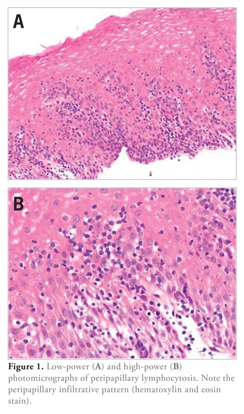
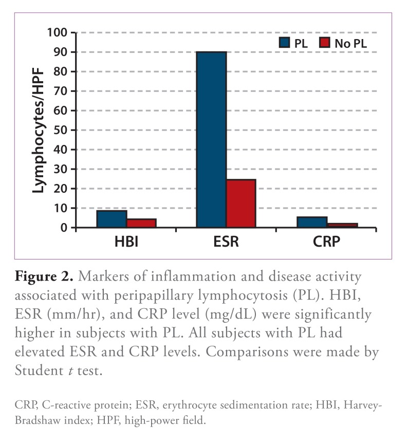
Lymphocytic esophagitis (LE) is a newly described entity characterized histopathologically by peripapillary lymphocytosis (PL) without significant granulocytes (neutrophils and eosinophils). In an initial study, a significant portion of patients with LE had Crohn's disease (CD). A subsequent study revealed LE in one quarter of children with CD. The aim of this study was to test the hypothesis that LE is associated with adult inflammatory bowel disease (IBD) and assess the disease variables that link LE and IBD. Random esophageal biopsies from consecutive adults with CD, ulcerative colitis (UC), or indeterminate colitis (IC) were evaluated. The numbers of lymphocytes, eosinophils, and neutrophils were counted from 3 high-power fields (HPF) in each specimen. Four of 47 patients (8.5%; 3/30 CD, 1/15 UC, and 0/2 IC) had PL (esophageal biopsies with ≥50 lymphocytes/HPF; mean, 100.5±31.1/HPF). A significant number of granulocytes were seen in biopsies from 3 of the 4 patients with PL, leaving 1 who met criteria for LE (PL without significant granulocytes). PL was associated with a higher erythrocyte sedimentation rate (90.3±17.6 mm/hr vs 24.5±3.6 mm/hr; P<.001) and C-reactive protein level (5.5±2.2 mg/dL vs 1.0±0.2 mg/dL; P<.001), with risk ratios of 2.06 (95% confidence interval [CI], 1.45-2.93; P=.031) and 3.56 (95% CI, 2.04-6.19; P=.033), respectively, for elevated values. All patients with PL had a relapsing CD course. The mean Harvey-Bradshaw index (HBI) was higher in these patients (8.5±0.6 vs 4.3±0.7; P=.026), with a risk ratio of 4.78 for moderate-to-severe disease (95% CI, 2.67-8.54; P=.004). We found a less frequent association between IBD and LE than was previously reported. This may be due to differences between pediatric and adult IBD. Alternatively, it may be methodologic because, unlike in previous reports, we evaluated consecutive patients with IBD. PL was associated with elevated inflammatory markers and HBI. These observations suggest that PL may be a marker of disease activity in IBD.
Keywords: Crohn’s disease; esophagitis; inflammatory bowel disease; lymphocytes; lymphocytic esophagitis; ulcerative colitis.
Figures
Similar articles
-
Lymphocytic esophagitis: a possible manifestation of pediatric upper gastrointestinal Crohn's disease.Inflamm Bowel Dis. 2011 Jan;17(1):45-9. doi: 10.1002/ibd.21347. Inflamm Bowel Dis. 2011. PMID: 20848529
-
Visceral adiposity and inflammatory bowel disease.Int J Colorectal Dis. 2021 Nov;36(11):2305-2319. doi: 10.1007/s00384-021-03968-w. Epub 2021 Jun 9. Int J Colorectal Dis. 2021. PMID: 34104989 Review.
-
Redefining the role of lymphocytes in gastroesophageal reflux disease and eosinophilic esophagitis.Dis Esophagus. 2010 Jul;23(5):368-76. doi: 10.1111/j.1442-2050.2010.01050.x. Epub 2010 Mar 26. Dis Esophagus. 2010. PMID: 20353445
-
CD4+ immune response as a potential biomarker of patient reported inflammatory bowel disease (IBD) activity.Clin Chim Acta. 2013 Jun 5;421:31-3. doi: 10.1016/j.cca.2013.02.016. Epub 2013 Feb 26. Clin Chim Acta. 2013. PMID: 23485644
-
Differentiating ulcerative colitis from Crohn disease in children and young adults: report of a working group of the North American Society for Pediatric Gastroenterology, Hepatology, and Nutrition and the Crohn's and Colitis Foundation of America.J Pediatr Gastroenterol Nutr. 2007 May;44(5):653-74. doi: 10.1097/MPG.0b013e31805563f3. J Pediatr Gastroenterol Nutr. 2007. PMID: 17460505
Cited by
-
Post-ablation lymphocytic esophagitis in Barrett esophagus with high grade dysplasia or intramucosal carcinoma.Mod Pathol. 2016 Jun;29(6):599-606. doi: 10.1038/modpathol.2016.50. Epub 2016 Mar 11. Mod Pathol. 2016. PMID: 26965580
-
Mucosal lesions of the upper gastrointestinal tract in patients with ulcerative colitis: A review.World J Gastroenterol. 2021 Jun 14;27(22):2963-2978. doi: 10.3748/wjg.v27.i22.2963. World J Gastroenterol. 2021. PMID: 34168401 Free PMC article. Review.
-
How to Approach Lymphocytic Esophagitis.Curr Gastroenterol Rep. 2017 Jun;19(6):24. doi: 10.1007/s11894-017-0564-y. Curr Gastroenterol Rep. 2017. PMID: 28429201 Review.
-
Clinical, endoscopic, and histologic characteristics of lymphocytic esophagitis: a systematic review.Esophagus. 2019 Apr;16(2):123-132. doi: 10.1007/s10388-018-0649-1. Epub 2018 Oct 29. Esophagus. 2019. PMID: 30370453
-
Esophagitis unrelated to reflux disease: current status and emerging diagnostic challenges.Virchows Arch. 2018 Jan;472(1):29-41. doi: 10.1007/s00428-017-2238-4. Epub 2017 Oct 15. Virchows Arch. 2018. PMID: 29034419 Review.
References
-
- Rubio CA, Sjodahl K, Lagergren J. Lymphocytic esophagitis: a histologic subset of chronic esophagitis. Am J Clin Pathol. 2006;125(3):432–437. - PubMed
-
- Rubio CA, Hubbard GB. Hyperplastic foveolar gastropathy and hyperplastic foveolar gastritis in baboons. In Vivo. 1996;10(5):507–510. - PubMed
-
- Ismail-Beigi F, Horton PF, Pope CE., 2nd Histological consequences of gastroesophageal reflux in man. Gastroenterology. 1970;58(2):163–174. - PubMed
-
- Black DD, Haggitt RC, Orenstein SR, Whitington PF. Esophagitis in infants: morphometric histological diagnosis and correlation with measures of gastroesophageal reflux. Gastroenterology. 1990;98(6):l408–l4l4. - PubMed
-
- Ismail-Beigi F, Pope CE., 2nd Distribution of the histological changes of gastroesophageal reflux in the distal esophagus of man. Gastroenterology. 1974;66(6):1109–1113. - PubMed
LinkOut - more resources
Full Text Sources
Research Materials
Miscellaneous