

Review
doi: 10.1155/2014/376367.
Epub 2014 Feb 26.
Endoscopic Optical Coherence Tomography (OCT): Advances in Gastrointestinal Imaging
Affiliations
- PMID: 24719611
- PMCID: PMC3955614
- DOI: 10.1155/2014/376367
Item in Clipboard
Review
Endoscopic Optical Coherence Tomography (OCT): Advances in Gastrointestinal Imaging
Gastroenterol Res Pract.
2014.
Abstract
In the rapidly evolving field of endoscopic gastrointestinal imaging, Optical Coherence Tomography (OCT) has found many diverse applications. We present the current status of OCT and its practical applications in imaging normal and abnormal mucosa in the esophagus, stomach, small and large intestines, and biliary and pancreatic ducts. We highlight technical aspects and principles of imaging, assess published data, and suggest future directions for OCT-guided evaluation and therapy.
Figures
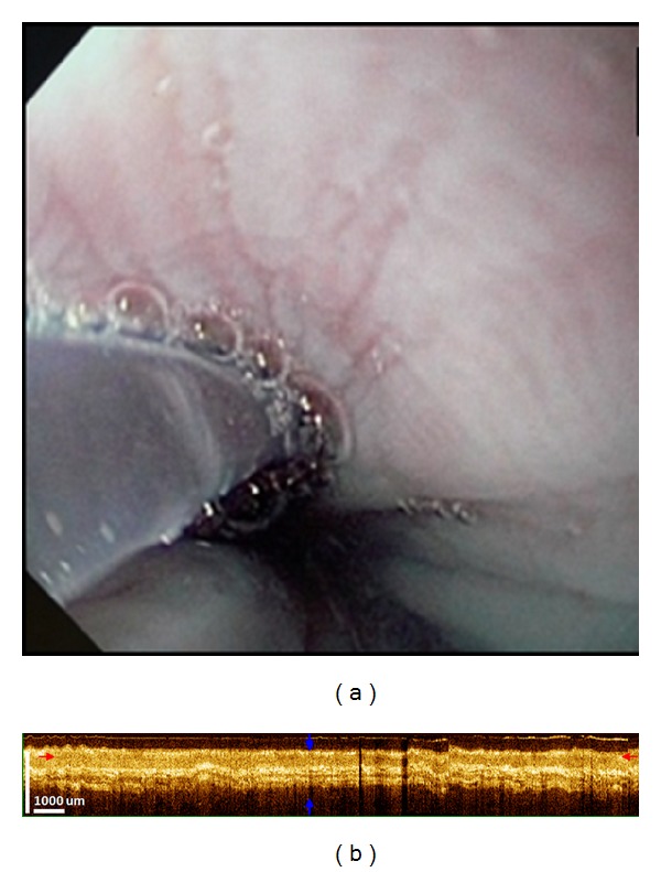
OCT imaging of normal esophagus. (a) Conventional endoscopy of the esophagus showing smooth pale mucosa. (b) Corresponding OCT image showing a well-defined, layered architecture. The epithelium, lamina propria, muscularis mucosa, submucosa, and muscularis propria are seen as distinct layers with alternating hypo- and hyperintensity.
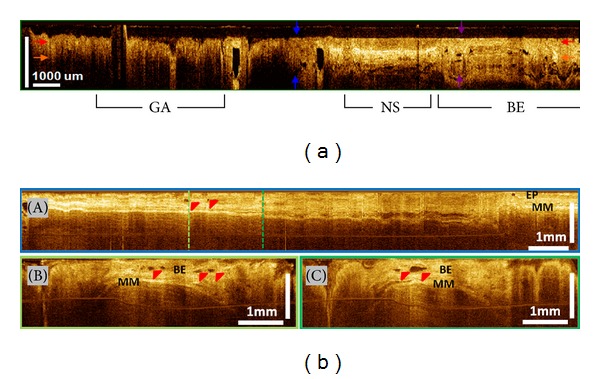
Barrett's esophagus (BE) without dysplasia. (a) Cross-sectional OCT imaging showing clear differences in layered architecture between gastric (GA), normal squamous (NS), and BE regions. BE regions exhibit distortion of the layered architecture and abnormal glandular features. (b) Cross-sectional OCT images around GEJ. BE glands (red arrows) are clearly observed (EP: epithelium; MM: muscularis mucosae in photos A–C).
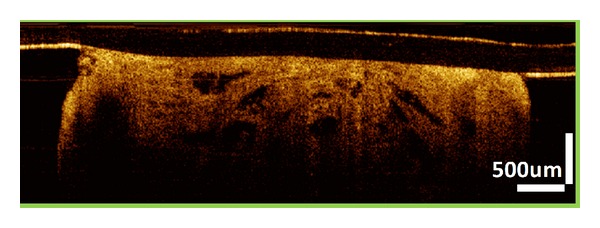
Intramucosal esophageal adenocarcinoma. OCT image showing dense large glands within the specimen. Lamina propria and muscularis mucosae (MM) layers are not clearly visible due to the infiltration of metaplasia into the MM layer.
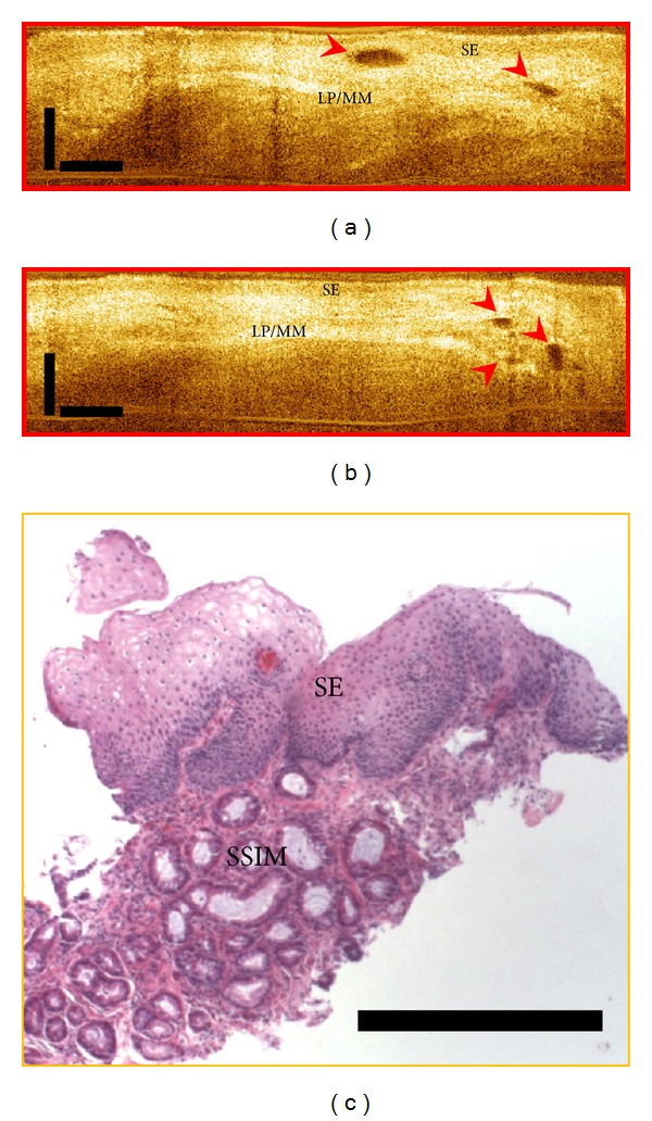
Subsquamous Intestinal Metaplasia (SSIM). (a-b): OCT images showing “buried glands” (red arrows). SE: squamous epithelium; LP/MM: lamina propria/muscularis mucosae. (c) Corresponding pathology showing subsquamous intestinal metaplasia under squamous epithelium.
References
-
- Song LM, Adler DG, Conway JD, et al. Narrow band imaging and multiband imaging. Gastrointestinal Endoscopy. 2008;67(4):581–589. - PubMed
-
- Neumann H, Kiesslich R, Wallace MB, Neurath MF. Confocal laser endomicroscopy: technical advances and clinical applications. Gastroenterology. 2010;139(2):388.e2–392.e2. - PubMed
-
- Tearney GJ, Brezinski ME, Southern JF, Bouma BE, Boppart SA, Fujimoto JG. Optical biopsy in human gastrointestinal tissue using optical coherence tomography. American Journal of Gastroenterology. 1997;92(10):1800–1804. - PubMed
Publication types
LinkOut - more resources
Full Text Sources
Other Literature Sources
