

Cardiovascular Magnetic Resonance Imaging of Myocardial Interstitial Expansion in Hypertrophic Cardiomyopathy
- PMID: 24719675
- PMCID: PMC3973947
- DOI: 10.1007/s12410-014-9267-z
Cardiovascular Magnetic Resonance Imaging of Myocardial Interstitial Expansion in Hypertrophic Cardiomyopathy
Abstract
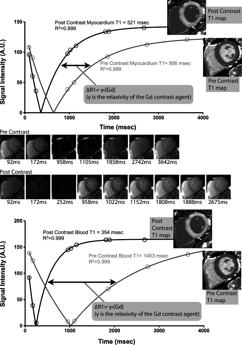
Hypertrophic cardiomyopathy (HCM) is a cardiovascular genetic disease with a varied clinical presentation and phenotype. Although mutations are typically found in genes coding for sarcomeric proteins, phenotypic derangements extend beyond the myocyte to include the extracellular compartment. Myocardial fibrosis is commonly detected by histology, and is associated with clinical vulnerability to adverse outcomes. Over the past decade, the noninvasive visualization of myocardial fibrosis by cardiovascular magnetic resonance (CMR) techniques has garnered much interest given the potential applications toward improving our understanding of pathophysiologic mechanisms of disease, as well as diagnosis and prognosis. Late gadolinium enhancement (LGE) imaging techniques are able to detect focal (typically replacement) fibrosis. Newer CMR techniques that measure absolute T1 relaxation time allow the quantification of the entire range of focal to diffuse (interstitial) fibrosis and may overcome potential limitations of LGE. This review will discuss the methodology and current status of these novel techniques, with a focus on extracellular volume fraction (ECV). Recent findings describing ECV measurement in HCM will be summarized.
Keywords: Cardiovascular magnetic resonance; Extracellular volume fraction; Gadolinium contrast; Hypertrophic cardiomyopathy; Myocardial fibrosis; T1 mapping.
Conflict of interest statement
Timothy C. Wong declares that he has no conflict of interest.
Figures
References
-
- Gersh BJ, Maron BJ, Bonow RO, et al. 2011 ACCF/AHA Guideline for the Diagnosis and Treatment of Hypertrophic Cardiomyopathy: a report of the American College of Cardiology Foundation/American Heart Association Task Force on Practice Guidelines. Circulation. 2011;124:e783–831. doi: 10.1161/CIR.0b013e318223e2bd. - DOI - PubMed
-
- Maron BJ, Gardin JM, Flack JM, Gidding SS, Kurosaki TT, Bild DE. Prevalence of hypertrophic cardiomyopathy in a general population of young adults. Echocardiographic analysis of 4111 subjects in the CARDIA Study. Coronary Artery Risk Development in (Young) Adults. Circulation. 1995;92:785–9. doi: 10.1161/01.CIR.92.4.785. - DOI - PubMed
Publication types
LinkOut - more resources
Full Text Sources
Other Literature Sources
Miscellaneous