

doi: 10.1155/2014/480573.
Epub 2014 Feb 25.
A clinical and pathological overview of vulvar condyloma acuminatum, intraepithelial neoplasia, and squamous cell carcinoma
Affiliations
- PMID: 24719870
- PMCID: PMC3956289
- DOI: 10.1155/2014/480573
Item in Clipboard
A clinical and pathological overview of vulvar condyloma acuminatum, intraepithelial neoplasia, and squamous cell carcinoma
Biomed Res Int.
2014.
Abstract
Condyloma acuminatum, intraepithelial neoplasia, and squamous cell carcinoma are three relatively frequent vulvar lesions. Condyloma acuminatum is induced by low risk genotypes of human papillomavirus (HPV). Vulvar intraepithelial neoplasia (VIN) and squamous cell carcinoma have different etiopathogenic pathways and are related or not with high risk HPV types. The goal of this paper is to review the main pathological and clinical features of these lesions. A special attention has been paid also to epidemiological data, pathological classification, and clinical implications of these diseases.
Figures
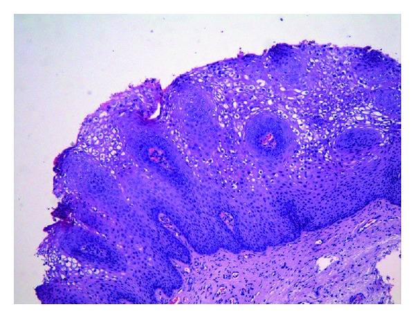
Vulvar condyloma acuminatum with acanthotic squamous epithelium and prominent koilocytic changes.
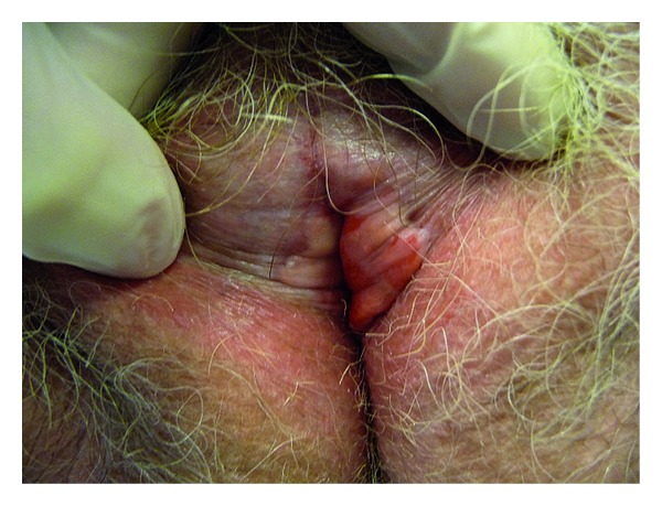
Differentiated VIN developed on sclerous lichen: white and surelevated nodules.
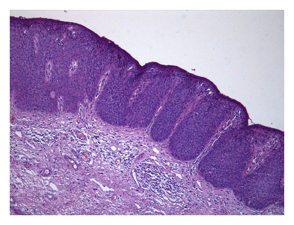
uVIN 3, basaloid type composed of a homogeneous population of dysplastic parabasal type cells on nearly whole thickness of the epidermis.
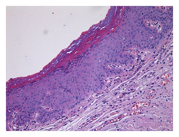
Differentiated VIN: atypical keratinocytes (with large vesicular nuclei with macronucleoli), present in the basal as well as mid layers of the epithelium. No koilocytic changes are identified.
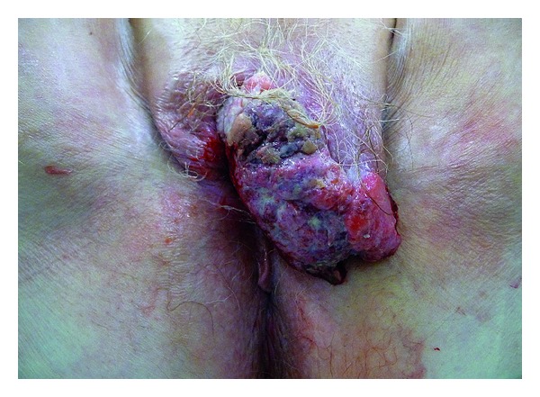
Exophytic and ulcerated squamous cell carcinoma.
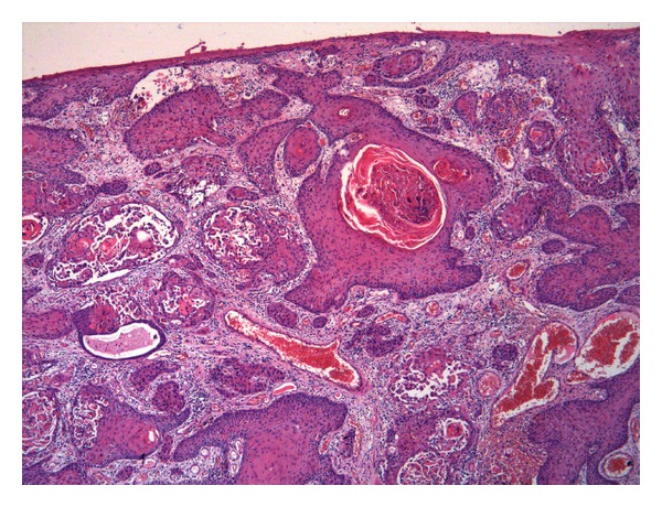
Keratinizing squamous cell carcinoma: infiltrative neoplastic cells are mature with abundant eosinophilic cytoplasm and show keratin pearls.
Similar articles
-
Coexisting high-grade vulvar intraepithelial neoplasia (VIN) and condyloma acuminatum: independent lesions due to different HPV types occurring in immunocompromised patients.Am J Surg Pathol. 2013 Jan;37(1):53-60. doi: 10.1097/PAS.0b013e318263cda6. Am J Surg Pathol. 2013. PMID: 23026935 Free PMC article.
-
Human papillomavirus infection and p16(INK4a) protein expression in vulvar intraepithelial neoplasia and invasive squamous cell carcinoma.J Low Genit Tract Dis. 2005 Apr;9(2):108-13. doi: 10.1097/00128360-200504000-00007. J Low Genit Tract Dis. 2005. PMID: 15870532
-
[Morphologic manifestations of human papillomavirus infection in the vulvar and anogenital region].Med Pregl. 1998 May-Jun;51(5-6):265-70. Med Pregl. 1998. PMID: 9720356 Croatian.
-
Pathways of vulvar intraepithelial neoplasia and squamous cell carcinoma.Histopathology. 2013 Jan;62(1):161-75. doi: 10.1111/his.12034. Epub 2012 Nov 27. Histopathology. 2013. PMID: 23190170 Review.
-
[HPV-associated alterations of the vulva and vagina. Morphology and molecular pathology].Pathologe. 2011 Nov;32(6):467-75. doi: 10.1007/s00292-011-1476-5. Pathologe. 2011. PMID: 22038133 Review. German.
Cited by
-
High-grade squamous intraepithelial lesion arising adjacent to vulvar lymphangioma circumscriptum: a tertiary institutional experience.Oncotarget. 2016 Jul 26;7(30):48120-48129. doi: 10.18632/oncotarget.10158. Oncotarget. 2016. PMID: 27329721 Free PMC article.
-
Squamous Cell Carcinoma In Situ-The Importance of Early Diagnosis in Bowen Disease, Vulvar Intraepithelial Neoplasia, Penile Intraepithelial Neoplasia, and Erythroplasia of Queyrat.Diagnostics (Basel). 2024 Aug 16;14(16):1799. doi: 10.3390/diagnostics14161799. Diagnostics (Basel). 2024. PMID: 39202286 Free PMC article. Review.
-
The effect of COVID-19 (SARS-CoV-2) vaccines on vulvar condylomata.J Turk Ger Gynecol Assoc. 2025 Jun 10;26(2):116-120. doi: 10.4274/jtgga.galenos.2025.2025-4-8. J Turk Ger Gynecol Assoc. 2025. PMID: 40495551 Free PMC article.
-
Cervical HPV infection in Guangzhou, China: an epidemiological study of 198,111 women from 2015 to 2021.Emerg Microbes Infect. 2023 Dec;12(1):e2176009. doi: 10.1080/22221751.2023.2176009. Emerg Microbes Infect. 2023. PMID: 36744409 Free PMC article.
-
Efficacy of 5% imiquimod cream on vulvar intraepithelial neoplasia in Korea: pilot study.Ann Dermatol. 2015 Feb;27(1):66-70. doi: 10.5021/ad.2015.27.1.66. Epub 2015 Feb 3. Ann Dermatol. 2015. PMID: 25673934 Free PMC article.
References
-
- Kurman RJ, Ellenson LH, Ronnett BM. Blaustein's Pathology of the Female Genital Tract. 6th edition. New York, NY, USA: Springer; 2011.
-
- Johansson H, Bzhalava D, Ekström J, Hultin E, Dillner J, Forslund O. Metagenomic sequencing of “HPV-negative” condylomas detects novel putative HPV types. Virology. 2013;440(1):1–7. - PubMed
-
- Cremin S, Menton JF, Canier L, Horgan M, Fanning LJ. The prevalence and genotype of human papillomavirus on cervical samples from an Irish female population with external genital warts. Human Vaccin & Immunotherapeutics. 2012;8(7):916–920. - PubMed
-
- Nucci MR, Oliva E. Gynecologic Pathology: A Volume in Foundation in Diagnostic Pathology Series. New York, NY, USA: Churchill Livingstone; 2009.
-
- Tavassoli FA, Devillée P. World Health Organization Classification of Tumours: Pathology and Genetics of Tumours of the Breast and Female Genital Organs. Lyon, France: IARC Press; 2003.
MeSH terms
LinkOut - more resources
Full Text Sources
Other Literature Sources
Medical
