

Sex differences in constitutive autophagy
- PMID: 24719882
- PMCID: PMC3955681
- DOI: 10.1155/2014/652817
Sex differences in constitutive autophagy
Abstract
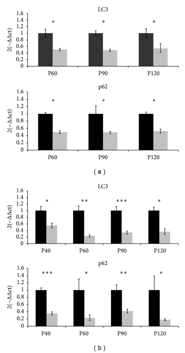
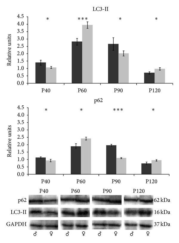
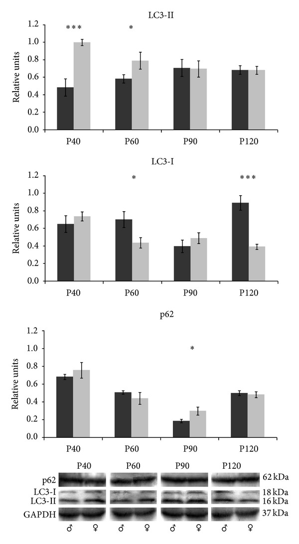
Sex bias has been described nowadays in biomedical research on animal models, although sexual dimorphism has been confirmed widely under pathological and physiological conditions. The main objective of our work was to study the sex differences in constitutive autophagy in spinal cord and skeletal muscle tissue from wild type mice. To examine the influence of sex on autophagy, mRNA and proteins were extracted from male and female mice tissues. The expressions of microtubule-associated protein 1 light chain 3 (LC3) and sequestosome 1 (p62), markers to monitor autophagy, were analyzed at 40, 60, 90, and 120 days of age. We found significant sex differences in the expression of LC3 and p62 in both tissues at these ages. The results indicated that sex and tissue specific differences exist in constitutive autophagy. These data underlined the need to include both sexes in the experimental groups to minimize any sex bias.
Figures
References
-
- Mizushima N, Komatsu M. Autophagy: renovation of cells and tissues. Cell. 2011;147(4):728–741. - PubMed
-
- Komatsu M, Waguri S, Chiba T, et al. Loss of autophagy in the central nervous system causes neurodegeneration in mice. Nature. 2006;441(7095):880–884. - PubMed
-
- Hara T, Nakamura K, Matsui M, et al. Suppression of basal autophagy in neural cells causes neurodegenerative disease in mice. Nature. 2006;441(7095):885–889. - PubMed
-
- Komatsu M, Ueno T, Waguri S, Uchiyama Y, Kominami E, Tanaka K. Constitutive autophagy: vital role in clearance of unfavorable proteins in neurons. Cell Death and Differentiation. 2007;14(5):887–894. - PubMed
Publication types
MeSH terms
LinkOut - more resources
Full Text Sources
Other Literature Sources
Research Materials
