Review
. 2014 Apr;171(8):1917-42.
doi: 10.1111/bph.12503.

Mitochondrial dysfunction and mitophagy: the beginning and end to diabetic nephropathy?

Affiliations
Review

Mitochondrial dysfunction and mitophagy: the beginning and end to diabetic nephropathy?

G C Higgins et al. Br J Pharmacol. 2014 Apr.

Abstract

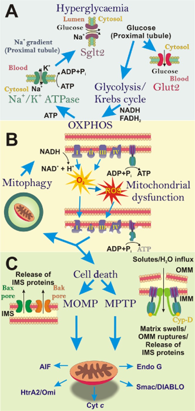
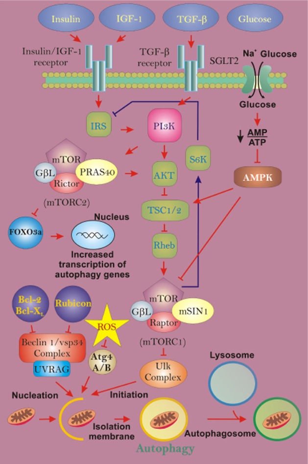
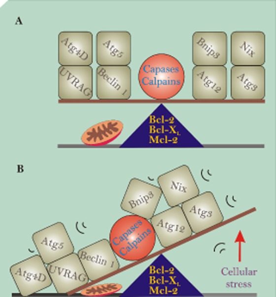
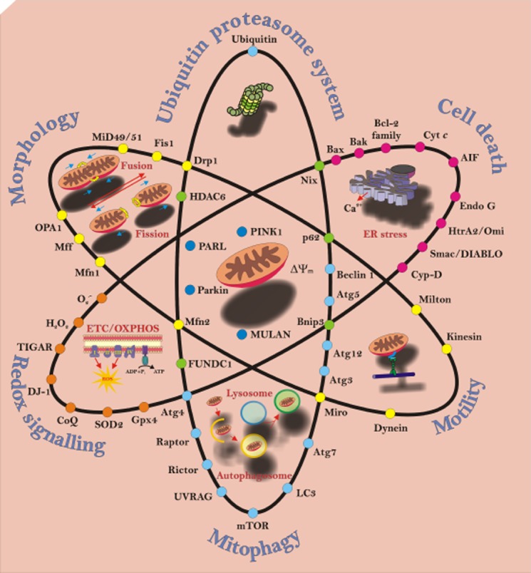
Diabetic nephropathy (DN) is a progressive microvascular complication arising from diabetes. Within the kidney, the glomeruli, tubules, vessels and interstitium are disrupted, ultimately impairing renal function and leading to end-stage renal disease (ESRD). Current pharmacological therapies used in individuals with DN do not prevent the inevitable progression to ESRD; therefore, new targets of therapy are urgently required. Studies from animal models indicate that disturbances in mitochondrial homeostasis are central to the pathogenesis of DN. Since renal proximal tubule cells rely on oxidative phosphorylation to provide adequate ATP for tubular reabsorption, an impairment of mitochondrial bioenergetics can result in renal functional decline. Defects at the level of the electron transport chain have long been established in DN, promoting electron leakage and formation of superoxide radicals, mediating microinflammation and contributing to the renal lesion. More recent studies suggest that mitochondrial-associated proteins may be directly involved in the pathogenesis of tubulointerstitial fibrosis and glomerulosclerosis. An accumulation of fragmented mitochondria are found in the renal cortex in both humans and animals with DN, suggesting that in tandem with a shift in dynamics, mitochondrial clearance mechanisms may be impaired. The process of mitophagy is the selective targeting of damaged or dysfunctional mitochondria to autophagosomes for degradation through the autophagy pathway. The current review explores the concept that an impairment in the mitophagy system leads to the accelerated progression of renal pathology. A better understanding of the cellular and molecular events that govern mitophagy and dynamics in DN may lead to improved therapeutic strategies.

Keywords: autophagy; diabetes; diabetic nephropathy; dynamics; kidney; mitochondria; mitochondrial dysfunction; mitophagy.


Figures

Figure 1
Figure 1
The ‘vicious cycle’ of diabetic nephropathy (DN): impact of hyperglycaemia on mitochondrial function. (A) In the diabetic milieu, excess blood glucose, filtered by the kidneys has to be reabsorbed, predominantly by the S1 segment of the proximal tubule. Glucose is transported through the proximal tubule epithelial cell (PTEC) via Sglt2 (on the apical/luminal surface) and GLUT2 (on the basolateral surface). Within the PTEC, this increased pool of glucose is metabolized and ATP is produced by oxidative phosphorylation (OXPHOS). The resultant ATP is utilized to fuel the Na+/K+-ATPase pump, which drives the Na+ gradient across the proximal tubule, required for co-transport of glucose via Sglt2. (B) During hyperglycaemia, superoxide (O2) is generated from the electron transport chain (ETC) and can be subsequently converted into other reactive oxygen species (ROS). Excess ROS causes damage to mitochondria and the ETC, resulting in impaired ATP production. These dysfunctional mitochondria can be eliminated by mitophagy. However, when the number of damaged mitochondria overwhelms the mitophagy process, dysfunctional mitochondria can initiate cell death. (C) Apoptosis or programmed necrosis ensues via mitochondria outer membrane permeabilization (MOMP) or mitochondrial permeability transition pore (MPTP). These mechanisms result in the release of inter-membrane space (IMS) proteins from mitochondria into the cytosol, which promote cell death.
Figure 2
Figure 2
Non-selective autophagy regulation during diabetic nephropathy (DN). During DN, autophagic turnover of mitochondria can be influenced by changes in several contributing factors, including insulin, IGF-1 (insulin-like growth factor-1), TGF-β, glucose and reactive oxygen species (ROS). These influences can act on the mechanistic target of rapamycin (mTOR) signalling pathway, which negatively regulates autophagy activity. Insulin, IGF-1 and TGF-β can modulate the mTOR pathway via the PI3K pathway. Increases in insulin or IGF-1 will suppress autophagy activity, while an increase in TGF-β will stimulate autophagy activity. Increases in glucose, with hyperglycaemia, can result in suppression of autophagy through activation of AMPK, which can activate the mTOR pathway. Finally, increases in ROS production can stimulate autophagy activation via inhibition of Atg4A/B or by directly acting on other components of the mTOR pathway, impairing activation.
Figure 3
Figure 3
Mitophagy players in life and death. (A) Depicts key autophagy proteins in perfect balance allowing the maintenance of mitochondrial homeostasis. (B) However, with increased cellular stress (e.g. glucose, reactive oxygen species), the balance can be tipped, resulting in some of these proteins being activated by proteases, such as caspases and calpains, whereas others interact with Bcl-2 family members through BH3-only domains.
Figure 4
Figure 4
Balancing mitochondrial homeostasis. This schematic illustrates the interaction between mitochondrial biogenesis, dynamics, organelle turnover and cell death, in addition to the major players involved in these processes. Mitochondria biogenesis involves oxidative phosphorylation (OXPHOS) via the electron transport chain (ETC). This process generates energy for the cell in the form of ATP, but in the process also generates reactive oxygen species (ROS). These ROS are involved in redox signalling regulated by antioxidants and transcription factors (orange circles). Mitochondria are dynamic organelles that are in constant motion, being transported around the cytoplasm while undergoing changing morphology (yellow circles). When mitochondria become dysfunctional via oxidative stress, mitochondrial dynamics is arrested and the ubiquitin proteasome system and mitophagy come into play to turnover the damaged organelles (pale blue circles). Central in regulating this process is the interaction between PARL (presenilin-associated rhomboid-like protease), PINK1 and Parkin (dark blue circles), and the involvement of adapter proteins (green circles). When cellular stress (e.g. damage to mitochondria and ER stress) overwhelms cellular homeostasis, cells can undergo diverse forms of death. Depending on the type and severity of the stress, mitochondria can regulate multiple cell death pathways involving several different signalling pathways (pink circles).

References

    1. The effect of intensive treatment of diabetes on the development and progression of long-term complications in insulin-dependent diabetes mellitus. The Diabetes Control and Complications Trial Research Group. N Engl J Med. 1993;329:977–986. - PubMed
    1. American Diabetes Association. Economic costs of diabetes in the U.S. in 2012. Diabetes Care. 2013;36:1033–1046. - PMC - PubMed
    1. Andrabi SA, Dawson TM, Dawson VL. Mitochondrial and nuclear cross talk in cell death: parthanatos. Ann N Y Acad Sci. 2008;1147:233–241. - PMC - PubMed
    1. Armstrong JS. The role of the mitochondrial permeability transition in cell death. Mitochondrion. 2006;6:225–234. - PubMed
    1. Atkinson SJ, Hosford MA, Molitoris BA. Mechanism of actin polymerization in cellular ATP depletion. J Biol Chem. 2004;279:5194–5199. - PubMed

Publication types

MeSH terms