. 2014 Apr 22;8(4):3251-9.
doi: 10.1021/nn4038223. Epub 2014 Apr 11.

Developmental self-assembly of a DNA tetrahedron

Affiliations

Developmental self-assembly of a DNA tetrahedron

John P Sadowski et al. ACS Nano. .

Abstract

Kinetically controlled isothermal growth is fundamental to biological development, yet it remains challenging to rationally design molecular systems that self-assemble isothermally into complex geometries via prescribed assembly and disassembly pathways. By exploiting the programmable chemistry of base pairing, sophisticated spatial and temporal control have been demonstrated in DNA self-assembly, but largely as separate pursuits. By integrating temporal with spatial control, here we demonstrate the "developmental" self-assembly of a DNA tetrahedron, where a prescriptive molecular program orchestrates the kinetic pathways by which DNA molecules isothermally self-assemble into a well-defined three-dimensional wireframe geometry. In this reaction, nine DNA reactants initially coexist metastably, but upon catalysis by a DNA initiator molecule, navigate 24 individually characterizable intermediate states via prescribed assembly pathways, organized both in series and in parallel, to arrive at the tetrahedral final product. In contrast to previous work on dynamic DNA nanotechnology, this developmental program coordinates growth of ringed substructures into a three-dimensional wireframe superstructure, taking a step toward the goal of kinetically controlled isothermal growth of complex three-dimensional geometries.


Figures
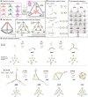
Figure 1
Figure 1
Catalytic self-assembly of a DNA tetrahedron. (a) Overview of the reaction. (b) A computer-rendered model of the tertiary structure of the assembled tetrahedron. (c) The reaction graph of the developmental molecular program specifying kinetically controlled self-assembly (left) compared to a traditional self-assembly process that lacks pathway control (right). Solid and dashed arrows depict kinetically controlled assembly and disassembly operations; line segments depict assembly operations that are not kinetically controlled. (d) Execution schematics of two elementary reactions. Left, molecular structures; right, corresponding nodal abstractions, where lines are added to connect ports once assembly has occurred. The strand regions are colored the same as the corresponding ports in the nodal representation. Top, a hairpin assembly reaction. Bottom, a cooperative assembly reaction. (e) Execution of the developmental molecular program along one possible assembly pathway. Only active toehold domains are labeled in this figure, with newly hybridized toeholds labeled in pink. Top, molecular structures; bottom, corresponding nodal execution schematics. See Supporting Information Figure S1 for a schematic showing all sequence domains. (f) The full set of intermediates along the prescribed assembly pathways. A three-letter code is used to identify each species as explained in the text. Species that are structurally congruent are linked by gray boxes. The numbers of assembled strands (excluding the initiator) for each row are displayed. The pathway in panel e corresponds to the reactions along the left edge of this figure.
Figure 2
Figure 2
Characterization of the tetrahedron assembly pathway. This is a gel electrophoresis mobility shift assay where lanes 1–25 show all on-pathway intermediates of the tetrahedron assembly, formed by mixing different subsets of reactants with the initiator. Gray boxes mark groups of intermediates that are structurally congruent and expected to have the same mobility, and the structures of the intended products are shown, as in Figure 1f. Lanes 26–35 show the analysis of catalytic turnover at two concentrations, showing reactions containing all nine reactants but varying concentrations of initiator I. These are 6% native polyacrylamide gels of assembly reactions containing 1 equiv of all reactants at the specified concentration, except A1 for which we used 0.9 equiv of a FAM fluorophore-labeled hairpin to observe incorporation yields. The initiator was included at 1 equiv unless otherwise specified in the figure. The assembly reactions were conducted at room temperature in TAE/Mg2+ buffer containing 12.5 mM Mg2+ over 19 h. The dotted line separates two gel slabs that were run simultaneously, and the solid line separates gels that were run at different times. The intensity of the FAM fluorescent label is shown in red, and SYBR Gold staining intensity is shown in blue; these channels are separated in Supporting Information Figure S2. See Supporting Information Figure S4 for an agarose gel of these same samples, in which the side products of higher molecular weight are well-resolved.
Figure 3
Figure 3
Characterization of ring-forming reactions. In this fluorescence-quenching assay, each of the three C strands was functionalized with a different fluorophore at its 5′ end (C1–TYE 665, red; C2–TAMRA, green; C3–FAM, blue); F represents a C strand with a fluorophore only, while Q represents a strand with a fluorophore expected to be quenched by a quencher on the 3′-end of a neighboring C strand. Lanes 1–3 show the structures FBB (CBB with a red fluorophore on the C1 strand), FFB (CCB with red and green fluorophores on the C1 and C2 strands, respectively), and QFB (CCB with the same two fluorophores, plus a 3′-quencher on the C2 strand that quenches the red 5′-fluorophore on the C1 strand). Lanes 4–6 and 7–9 show the other two structural permutations. Lane 10 shows FFF (the full tetrahedron with all fluorophores), and lane 11 shows QQQ (the full tetrahedron with all fluorophores and all quenchers). Lanes 12 and 13 are the same lanes in the same gel as lanes 10 and 11, but after staining with SYBR Gold. This is a 6% native polyacrylamide gel of a 10 nM assembly reaction with 1 equiv of initiator, conducted at room temperature in TAE/Mg2+ buffer containing 12.5 mM Mg2+ over 22 h.
Figure 4
Figure 4
Atomic force microscopy images of tetrahedron self-assembly. (a) Images of purified samples of the intermediates BBB, CBB, CCB, and the full tetrahedron CCC, which, respectively, appeared as a three-arm junction, a single triangle, a double triangle, and a three- or four-lobed structure corresponding to a flattened tetrahedron, each consistent with our design. In each image, the double-stranded edges of each wireframe structure are clearly resolved. (b) Images of the full tetrahedron cut by restriction endonucleases followed by incubation with streptavidin. Digestion with either of two endonucleases targeted to different edges restored the double-triangle structures, with the streptavidin (appearing as small white circular features in the AFM image) appearing at the expected biotin-modified vertices. In the schematic diagrams, the black bar intersecting tetrahedron edge represents the designed restriction site, the black dot is the 5′-biotin, and the yellow circle is the streptavidin. The scale bars of the larger images are 100 nm long; the inset images are 62.5 nm in width, at double the scale of the larger images. These samples were assembled at a concentration of 100 nM and then purified by glycerol gradient ultracentrifugation; for panel b, the restriction and streptavidin binding were performed before purification.

References

    1. Seeman N. C. Nucleic Acid Junctions and Lattices. J. Theor. Biol. 1982, 99, 237–247. - PubMed
    1. Seeman N. C. DNA in a Material World. Nature 2003, 421, 427–431. - PubMed
    1. Shih W. M.; Lin C. Knitting Complex Weaves with DNA Origami. Curr. Opin. Struct. Biol. 2010, 20, 276–282. - PMC - PubMed
    1. Schulman R.; Winfree E. Synthesis of Crystals with a Programmable Kinetic Barrier to Nucleation. Proc. Natl. Acad. Sci. U.S.A. 2007, 104, 15236–15241. - PMC - PubMed
    1. Yin P.; Hariadi R.; Sahu S.; Choi H. M. T.; Park S. H.; LaBean T. H.; Reif J. H. Programming Molecular Tube Circumferences. Science 2008, 321, 824–826. - PubMed

Publication types