

Video-mosaicing of reflectance confocal images for examination of extended areas of skin in vivo
- PMID: 24720744
- PMCID: PMC4405240
- DOI: 10.1111/bjd.13050
Video-mosaicing of reflectance confocal images for examination of extended areas of skin in vivo
Abstract
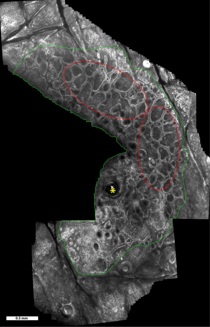
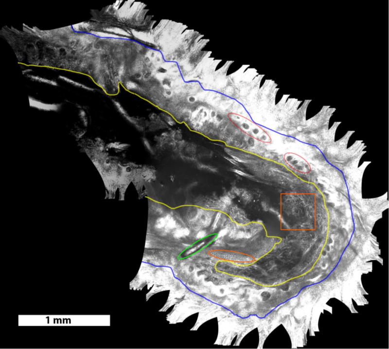
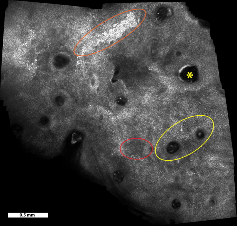
Background: With reflectance confocal microscopy (RCM) imaging, skin cancers can be diagnosed in vivo and margins detected to guide treatment. Since the field of view of an RCM image is much smaller than the typical size of lesions, mosaicing approaches have been developed to display larger areas of skin. However, the current paradigm for RCM mosaicing in vivo is limited both in speed and to pre-selected rectangular-shaped small areas. Another approach, called “video-mosaicing,” enables higher speeds and real-time operator-selected areas of any size and shape, and will be more useful for RCM examination of skin in vivo.
Objectives: To demonstrate the feasibility and clinical potential of video-mosaicing of RCM images to rapidly display large areas of skin in vivo.
Methods: Thirteen videos of benign lesions, melanocytic cancers and residual basal cell carcinoma margins were collected on volunteer subjects with a handheld RCM scanner. The images from each video were processed and stitched into mosaics to display the entire area that was imaged.
Results: Acquisition of RCM videos covering 5.0–16.0 mm2 was performed in 20–60 seconds. The video-mosaics were visually determined to be of high quality for resolution, contrast and seamless contiguity, and the appearance of cellular-level and morphologic detail.
Conclusion: Video-mosaicing confocal microscopy, with real-time operator-choice of the shape and size of the area to be imaged, will enable rapid examination of large areas of skin in vivo. This approach may further advance noninvasive detection of skin cancer and, eventually, facilitate wider adoption of RCM imaging in the clinic.
Figures
References
-
- Guitera P, Menzies SW, Longo C, Cesinaro AM, Scolyer RA, Pellacani G. In Vivo Confocal Microscopy for Diagnosis of Melanoma and Basal Cell Carcinoma Using a Two-Step Method: Analysis of 710 Consecutive Clinically Equivocal Cases. Journal of Investigative Dermatology. 2012;132:2386–2394. - PubMed
-
- Guitera P, Pellacani G, Crotty KA, Scolyer RA, Li LX, Bassoli S, Vinceti M, Rabinovitz H, Longo C, Menzies SW. The impact of in vivo reflectance confocal microscopy on the diagnostic accuracy of lentigo maligna and equivocal pigmented and nonpigmented macules of the face. 2010;130(8):2080–91. - PubMed
-
- Guitera P, Moloney FJ, Menzies SW, Stretch J, Quinn MJ, Hong A, Fogarty G, Scolyer RA. Improving management and patient care of lentigo maligna by mapping with in vivo confocal microscopy. JAMA Dermatology. 2013;149(6):692–698. - PubMed
-
- Nadiminti H, Scope A, Marghoob AA, Busam K, Nehal KS. Use of Reflectance Confocal Microscopy to Monitor Response of Lentigo Maligna to Nonsurgical Treatment. Dermatologic Surgery. 2010;36(2):177–184. - PubMed
Publication types
MeSH terms
Grants and funding
LinkOut - more resources
Full Text Sources
Other Literature Sources
Medical
