. 2014 Jun;18(6):1113-23.
doi: 10.1111/jcmm.12256. Epub 2014 Apr 10.

Retinoic acid reduces migration of human breast cancer cells: role of retinoic acid receptor beta

Affiliations

Retinoic acid reduces migration of human breast cancer cells: role of retinoic acid receptor beta

Marina Ines Flamini et al. J Cell Mol Med. 2014 Jun.

Abstract

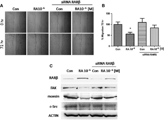
Breast cancer is the most common malignancy in women and the appearance of distant metastases produces the death in 98% of cases. The retinoic acid receptor β (RARβ) is not expressed in 50% of invasive breast carcinoma compared with normal tissue and it has been associated with lymph node metastasis. Our hypothesis is that RARβ protein participates in the metastatic process. T47D and MCF7 breast cancer cell lines were used to perform viability assay, immunobloting, migration assays, RNA interference and immunofluorescence. Administration of retinoic acid (RA) in breast cancer cells induced RARβ gene expression that was greatest after 72 hrs with a concentration 1 μM. High concentrations of RA increased the expression of RARβ causing an inhibition of the 60% in cell migration and significantly decreased the expression of migration-related proteins [moesin, c-Src and focal adhesion kinase (FAK)]. The treatment with RARα and RARγ agonists did not affect the cell migration. On the contrary, the addition of the selective retinoid RARβ-agonist (BMS453) significantly reduced cell migration comparable to RA inhibition. When RARβ gene silencing was performed, the RA failed to significantly inhibit migration and resulted ineffective to reduce moesin, c-Src and FAK expressions. RARβ is necessary to inhibit migration induced by RA in breast cancer cells modulating the expression of proteins involved in cell migration.

Keywords: FAK; RARβ; breast cancer cells; cell migration; moesin; retinoic acid.


Figures

Figure 1
Figure 1
(A and B) The cellular lines MCF7 and T47D were seeded at 13,000 cells per well and treated with RA (10−7/10−5 M) for 3 days. The results were expressed as percentage (%) of survival cells. All data shown were representative of three independent experiments. Error bars indicate standard deviations. *P < 0.05 versus Con.
Figure 2
Figure 2
(A) MCF7 cells were treated with retinoic acid (RA) in different concentrations (10−7/10−5 M) and cell migration was imaged after 72 hrs. (B) Gap closure was quantified with the use of NIH image J software. *P < 0.05 versus Con. (C) T47D cells were treated with RA (10−6 M) and the synthetic agonist retinoids, selective for RARα Agonist (BMS753), RARβ Agonist (BMS453) and RARγ Agonist (BMS961), and the synthetic antagonist retinoids selective for RARα (BMS195614) plus RA (10−6 M). All retinoids were incubated at 10−6 M for 72 hrs. Cell migration was imaged after 72 hrs. (D) Gap closure was quantified with the use of NIH image J software. *P < 0.05 versus Con. These experiments were performed in triplicates and representative images are shown.
Figure 3
Figure 3
(A) Western blot analysis for RARβ protein was performed in 30 μg of total lysate of untreated MDA-MB231, T47D and MCF7 breast cancer cells. (B) Western blots show total cell amount of RARβ in MCF7 treated with different concentrations of retinoic acid (RA; 10−8/10−5 M) for 48 hrs. (C) MCF7 breast cancer cells were treated with RA (10−6 M) for 24, 48, 72 hrs and the RARβ, FAK, moesin, and c-Src expression are shown. Actin expression is shown in the lower boxes as loading control. The experiments were repeated three times with consistent results and representative images are shown. Densitometric quantifications of all the blots (including those not shown) were performed and the relative mean ± SD of each condition are presented in graph as supplemental data online Fig. S1A–C.
Figure 4
Figure 4
MCF7 cells were exposed to retinoic acid (RA; 10−6 M/72 hr) and another group was first transfected with a specific RARβ siRNA and then treated or not with RA (10−6 M/72 hr). (A) Cell migration was imaged after 72 hrs. (B) Gap closure was quantified with the use of NIH image J software. *P < 0.05 versus Con. (C) Western blot analysis for RARβ, FAK, moesin and c-Src. Actin expression is shown in the lower boxes as loading control. These experiments were performed in triplicates and representative images are shown. Densitometric quantifications of all the blots (including those not shown) were performed and the relative mean ± SD of each condition are presented in graph as supplemental data online Fig. S2.
Figure 5
Figure 5
T47D cells were exposed to retinoic acid (RA; 10−6 M/72 hr) and another group was first transfected with a control siRNA or specific RARβ siRNA and then treated or not with RA (10−6 M/72 hr). (A) Cell migration was imaged after 72 hrs. (B) Gap closure was quantified with the use of NIH image J software. *P < 0.05 versus Con. (C) Western blot analysis for RARβ, FAK, moesin, c-Src and Actin as loading control was performed. (D) Representative images of T47D cell adhesion to gelatin after RA treatment (10−6/10−5 M) for 72 hrs are shown. These experiments were performed in triplicates and representative images are shown. Densitometric quantifications of all the blots (including those not shown) were performed and the relative mean ± SD of each condition are presented in graph as supplemental data online Fig. S3.
Figure 6
Figure 6
(A and B) MCF7 and T47D cells were exposed to retinoic acid (RA; 10−6 M/72 hr) and another group was first transfected with a specific RARβ siRNA and then treated or not with RA (10−6 M/72 hr). Then the cells were stained with anti-moesin (clone 38) linked to Alexa Fluor 488 (green), actin was stained with phalloidin linked to Texas Red (red) and nuclei were counterstained with DAPI (blue). All the experiments were repeated three times with consistent results, and the representative images are shown. The microphotographs were taken with a 100 × objective.
Figure 7
Figure 7
(A) Immunofluorescence images of MCF7 and T47D cells stained with Texas Red-phalloidin (actin) were used to quantify the effects, of cells with a static and a migratory phenotype. The results were expressed as percentage (%) of Cells with Migratory Phenotype. (B) Representative images of Static and Migratory Phenotype of T47D cells are shown. All the experiments were repeated three times with consistent results, and the representative images are shown. The microphotographs were taken with a 100 × objective.

References

    1. Sporn MB. The war on cancer. Lancet. 1996;347:1377–81. - PubMed
    1. Lotan R, Lotan D, Sacks PG. Inhibition of tumor cell growth by retinoids. Methods Enzymol. 1990;190:100–10. - PubMed
    1. Ross SA, McCaffery PJ, Drager UC, et al. Retinoids in embryonal development. Physiol Rev. 2000;80:1021–54. - PubMed
    1. Connolly RM, Nguyen NK, Sukumar S. Molecular pathways: current role and future directions of the retinoic Acid pathway in cancer prevention and treatment. Clin Cancer Res. 2013;19:1651–9. - PMC - PubMed
    1. Chambon P. A decade of molecular biology of retinoic acid receptors. FASEB J. 1996;10:940–54. - PubMed

Publication types

MeSH terms