. 2014 Apr 9;15(4):471-83.
doi: 10.1016/j.chom.2014.03.003.

Neutrophils recruited by IL-22 in peripheral tissues function as TRAIL-dependent antiviral effectors against MCMV

Affiliations

Neutrophils recruited by IL-22 in peripheral tissues function as TRAIL-dependent antiviral effectors against MCMV

Maria A Stacey et al. Cell Host Microbe. .

Abstract

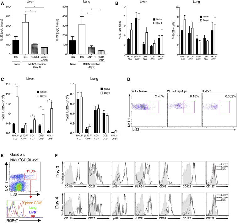
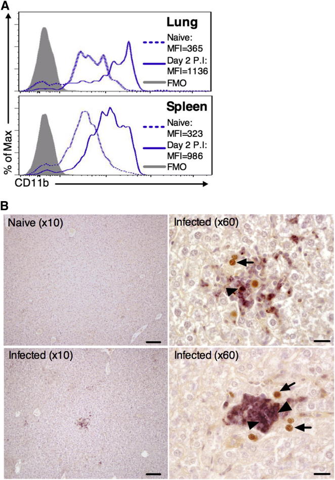
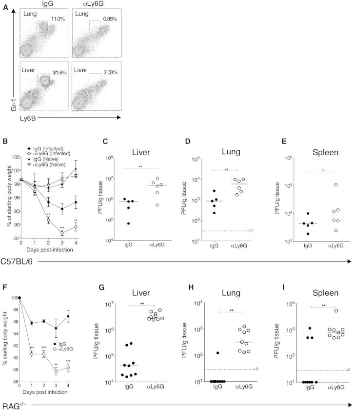
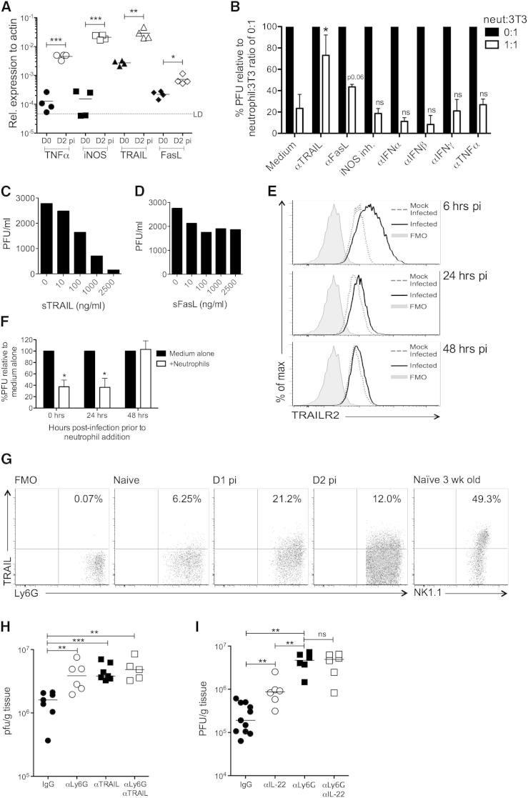
During primary infection, murine cytomegalovirus (MCMV) spreads systemically, resulting in virus replication and pathology in multiple organs. This disseminated infection is ultimately controlled, but the underlying immune defense mechanisms are unclear. Investigating the role of the cytokine IL-22 in MCMV infection, we discovered an unanticipated function for neutrophils as potent antiviral effector cells that restrict viral replication and associated pathogenesis in peripheral organs. NK-, NKT-, and T cell-secreted IL-22 orchestrated antiviral neutrophil-mediated responses via induction in stromal nonhematopoietic tissue of the neutrophil-recruiting chemokine CXCL1. The antiviral effector properties of infiltrating neutrophils were directly linked to the expression of TNF-related apoptosis-inducing ligand (TRAIL). Our data identify a role for neutrophils in antiviral defense, and establish a functional link between IL-22 and the control of antiviral neutrophil responses that prevents pathogenic herpesvirus infection in peripheral organs.


Figures

None
Graphical abstract
Figure 1
Figure 1
IL-22 Limits Acute MCMV Replication in a Tissue-Restricted Manner (A) IL-22R gene expression in the liver, lungs, and spleens of naive (day 0) and MCMV-infected mice day 2 p.i. (B–D) Replicating virus in livers (B), lungs (C), and spleens (D) of mice infected for 2 (triangles) and 4 (circles) days and treated with IgG or αIL-22. Results are expressed as PFU/g tissue. Individual mice + median values are shown. Dotted line, limit of detection (LD). (E) Weight loss is expressed as mean ± SEM (6 mice/group) of percent of starting weight. Data represent two to six experiments. See also Figure S1.
Figure 2
Figure 2
Conventional NK Cells, NK T Cells, and αβ T Cells Express IL-22 during MCMV Infection (A–C) Mice were infected with MCMV or mock infected (naive), and day 4 p.i. liver (left) and lung (right) tissue was isolated. (A) IL-22 protein concentrations within organ homogenates from naive mice, MCMV-infected mice (day 4 p.i.) treated with IgG, αNK1.1, or αCD4 and αCD8. (B and C) The proportion (B) and total numbers (C) of IL-22+ cells in naive and infected (day 4 p.i.) organs were assessed by FACS. Results show mean ± SEM of four to seven mice/group and represent two (A) or six (B and C) experiments studying expression at either day 2 or day 4 p.i. (D) Representative bivariate flow cytometry plots of IL-22 versus NK1.1 expression by hepatic NK1.1+CD3+ cells from WT (left and middle) or IL-22−/− mice day 0 or day 4 p.i., measured after 4 hr stimulation with PMA/ionomycin. (E) Expression of RORγT by NK1.1+CD3IL-22+ cells derived from the Peyer’s patch (PP), liver and lung, and splenic CD3+ cells from naive mice. (F) Representative histogram overlays of surface marker expression by IL-22+ (solid line) and IL-22 (dotted line) pulmonary NK cells day 0 and day 4 p.i. Results represent ≥3 experiments. See also Figure S2.
Figure 3
Figure 3
IL-22 Neutralization Impairs Neutrophil Recruitment into MCMV-Infected Tissues MCMV-infected mice were administered IgG or αIL-22 and leukocyte infiltrates (A–F) and chemokine protein expression (K–M) assessed after 2 days. Myeloid cell numbers in liver (A), lung (B), and spleen (C) were quantified and are shown as mean ± SEM of six mice per group. Representative bivariate plots of Ly6G+CD11b+ neutrophils in the liver (D), lung (E), and spleen (F). Results represent three experiments. (G–I) MCMV-infected mice were treated with IgG or αCXCL1 and neutrophil (Ly6G+CD11b+) recruitment into the liver (G), lung (H), and spleen (I) assessed by FACS. Results are expressed as mean + SEM of 11 mice, with data from two independent experiments combined. (J) 3T3 (IL-22R) and SGC1 (IL-22R+) cells were incubated for 6 hr in medium alone, 50 ng/ml rIL-22, or medium alone prior to MCMV infection (moi 0.5). Supernatants were then assayed for CXCL1 protein. Data are shown as mean + SEM of duplicate samples, representing four experiments. (K–M) CXCL1 protein in liver (K), lung (L), and spleen (M) homogenates from IgG- or αIL-22-treated mice day 2 p.i. Mean + SEM of eight (naive) or ten (MCMV infected ± αIL-22) mice is shown. Results are combined from two independent experiments, representing four in total.
Figure 4
Figure 4
Neutrophil Recruitment into MCMV-Infected Organs (A) CD11b expression by pulmonary (top) and splenic (bottom) neutrophils in infected (day 2 p.i.) or mock-infected mice. (B) Neutrophil localization near MCMV-infected (m123+) cells in the liver was visualized in hematoxylin-stained paraffin-embedded sections. Liver sections from mock-infected tissues (10×, top left) were used as negative controls for αm123 (brown) and αLy6G (purple) staining. Infected tissue is shown at 10× magnification (bottom left) and 60× (top and bottom right). All images are taken from different mice, representing <8. Arrow, MCMV-infected m123+ nucleus; triangle, neutrophil. Bars, 100 μM (10× magnification), 20 μM (60× magnification).
Figure 5
Figure 5
Neutrophil Depletion Impairs Control of Acute MCMV Infection MCMV-infected and mock-infected C57BL/6 (A–E) or RAG1−/− (F–I) mice were treated with αLy6G or IgG. (A) Representative bivariate plots demonstrating specific depletion of neutrophils (Ly6BintGr1hi) in lungs and livers with αLy6G antibody. (B) Weight loss in MCMV-infected and mock-infected IgG- and αLy6G-treated mice is expressed as percentage of original weight and is shown as mean ± SEM of ten mice per infected group and three mice per naive group. Replicating virus in livers (C), lungs (D), and spleens (E) 4 days p.i. in IgG- and αLy6G-treated mice is shown as individual mice + median. All data represent five independent experiments. (F–I) MCMV-infected RAG1−/− mice were treated with IgG or αLy6G. (F) Weight loss is expressed as percentage of original weight and is shown as mean ± SEM of five mice per group, representing two experiments. (G–I) Replicating virus in homogenates from the livers (G), lungs (H), and spleens (I) 4 days p.i. in IgG- and anti-Ly6G-treated RAG−/− mice. Individual mice + median from two experiments are shown.
Figure 6
Figure 6
Neutrophils Limit MCMV Replication In Vitro (A) MCMV-infected mice were treated ± αLy6G ± αNK1.1, and day 4 p.i. virus load was measured by plaque assay. Data from two experiments + median are shown. (B–D) Neutrophil purity was assessed by FACS (B), and survival following incubation with mock-infected and MCMV-infected fibroblasts was assessed with Annexin V and live/dead aqua (C). (D) Replicating virus in supernatants from neutrophil/fibroblast cocultures was measured after 7 days. Some wells received freeze-thawed neutrophils as negative controls. Data represent three (A), two (C), and four (B and D) experiments. See also Figure S3.
Figure 7
Figure 7
Neutrophils Limit MCMV Replication in a TRAIL-Dependent Manner (A) Whole-tissue gene expression in naive and infected (day 2 p.i.) livers. (B) Purified neutrophils were added or not at a ratio of 1:1 neutrophils:infected fibroblast ± antagonists of potential antiviral effector molecules. Relative reduction in MCMV PFU was compared between medium control and experimental groups. (C and D) Infected fibroblasts were incubated with recombinant TRAIL (C) or recombinant FasL (D) and replicating virus measured after 7 days. (E) Fibroblasts were MCMV infected (moi 0.2), and TRAILR expression was measured by FACS. Shaded, FMO; dotted lines, mock infected; solid line, MCMV infected. (F) Purified neutrophils were added or not to MCMV-infected fibroblasts 0, 24, or 48 hr p.i. and virus measured 7 days later. Data are shown as mean + SEM of four replicates. (G) Representative bivariant FACS plots of surface TRAIL expression by liver neutrophils. FMO, fluorescence minus one; positive control, NK1.1+CD3 cells from naive livers of 3-week-old mice. (H) MCMV-infected RAG−/− mice were depleted of NK1.1 cells and administered IgG (●), αLy6G (○), αTRAIL (▪) or αLy6G and αTRAIL (□), and hepatic virus load was measured at day 4 p.i. (I) MCMV-infected C57BL/6 mice were administered IgG (●), αIL-22 (○), αLy6G (▪), or αIL-22 and αLy6G (□) and virus load in liver tissue measured 4 days p.i. Results represent two (A, C, D, F, and G) or three (B and E) independent experiments, or show data merged from two experiments (H and I). See also Figure S4.

References

    1. Andoniou C.E., van Dommelen S.L., Voigt V., Andrews D.M., Brizard G., Asselin-Paturel C., Delale T., Stacey K.J., Trinchieri G., Degli-Esposti M.A. Interaction between conventional dendritic cells and natural killer cells is integral to the activation of effective antiviral immunity. Nat. Immunol. 2005;6:1011–1019. - PubMed
    1. Andrews D.M., Scalzo A.A., Yokoyama W.M., Smyth M.J., Degli-Esposti M.A. Functional interactions between dendritic cells and NK cells during viral infection. Nat. Immunol. 2003;4:175–181. - PubMed
    1. Aujla S.J., Chan Y.R., Zheng M., Fei M., Askew D.J., Pociask D.A., Reinhart T.A., McAllister F., Edeal J., Gaus K. IL-22 mediates mucosal host defense against Gram-negative bacterial pneumonia. Nat. Med. 2008;14:275–281. - PMC - PubMed
    1. Bai F., Kong K.F., Dai J., Qian F., Zhang L., Brown C.R., Fikrig E., Montgomery R.R. A paradoxical role for neutrophils in the pathogenesis of West Nile virus. J. Infect. Dis. 2010;202:1804–1812. - PMC - PubMed
    1. Banks T.A., Rickert S., Benedict C.A., Ma L., Ko M., Meier J., Ha W., Schneider K., Granger S.W., Turovskaya O. A lymphotoxin-IFN-beta axis essential for lymphocyte survival revealed during cytomegalovirus infection. J. Immunol. 2005;174:7217–7225. - PubMed

Publication types

LinkOut - more resources