

Maternal obesity and diabetes may cause DNA methylation alteration in the spermatozoa of offspring in mice
- PMID: 24721882
- PMCID: PMC3984639
- DOI: 10.1186/1477-7827-12-29
Maternal obesity and diabetes may cause DNA methylation alteration in the spermatozoa of offspring in mice
Abstract
Background: The adverse effects on offspring of diabetic and/or obese mothers can be passed to the next generation. However, the mechanisms behind this are still unclear. Epigenetics may play a key role during this process.
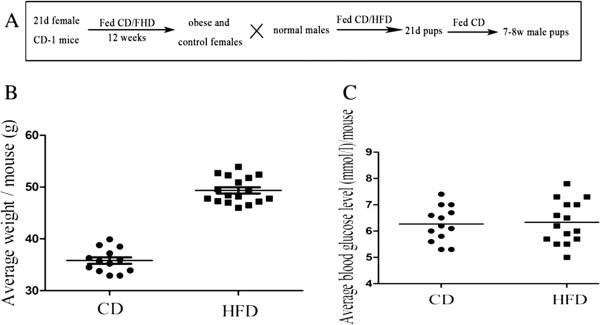
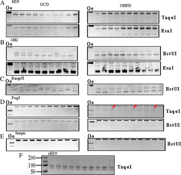
Methods: To confirm the hypothesis, we investigated the DNA methylation of several imprinted genes in spermatozoa of offspring from diabetic and/or obese mothers utilizing streptozotocin (STZ)- and high-fat-diet (HFD)-induced mouse models.
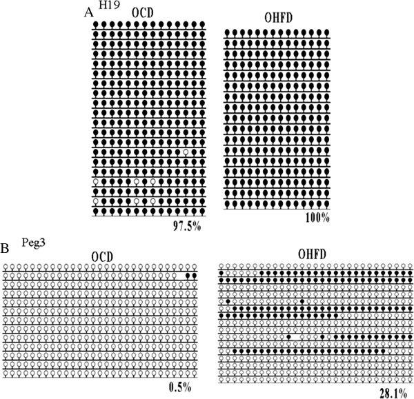
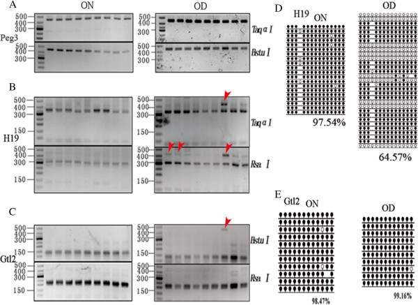
Results: We found that the DNA methylation of Peg3 was significantly increased in spermatozoa of offspring of obese mothers compared to that in spermatozoa of offspring of normal mothers. The DNA methylation of H19 was significantly higher in spermatozoa of offspring of diabetic mothers than that in spermatozoa of offspring of non-diabetic mothers.
Conclusions: These results indicate that pre-gestational diabetes and/or obesity can alter DNA methylation in offspring spermatozoa.
Figures
Similar articles
-
Intergenerational impact of paternal lifetime exposures to both folic acid deficiency and supplementation on reproductive outcomes and imprinted gene methylation.Mol Hum Reprod. 2017 Jul 1;23(7):461-477. doi: 10.1093/molehr/gax029. Mol Hum Reprod. 2017. PMID: 28535307 Free PMC article.
-
Maternal diabetes causes alterations of DNA methylation statuses of some imprinted genes in murine oocytes.Biol Reprod. 2013 May 9;88(5):117. doi: 10.1095/biolreprod.112.105981. Print 2013 May. Biol Reprod. 2013. PMID: 23515675
-
Maternal obesogenic diet combined with postnatal exposure to high-fat diet induces metabolic alterations in offspring.J Cell Physiol. 2020 Nov;235(11):8260-8269. doi: 10.1002/jcp.29482. Epub 2020 Jan 22. J Cell Physiol. 2020. PMID: 31970793
-
Aberrant DNA Methylation Mediates the Transgenerational Risk of Metabolic and Chronic Disease Due to Maternal Obesity and Overnutrition.Genes (Basel). 2021 Oct 20;12(11):1653. doi: 10.3390/genes12111653. Genes (Basel). 2021. PMID: 34828259 Free PMC article. Review.
-
Maternal diabetes in pregnancy: early and long-term outcomes on the offspring and the concept of "metabolic memory".Exp Diabetes Res. 2011;2011:218598. doi: 10.1155/2011/218598. Epub 2011 Nov 21. Exp Diabetes Res. 2011. PMID: 22144985 Free PMC article. Review.
Cited by
-
Sperm epigenetics and sperm RNAs as drivers of male infertility: truth or myth?Mol Cell Biochem. 2025 Feb;480(2):659-682. doi: 10.1007/s11010-024-04962-w. Epub 2024 May 8. Mol Cell Biochem. 2025. PMID: 38717684 Free PMC article. Review.
-
Long non-coding RNA H19 induces hippocampal neuronal apoptosis via Wnt signaling in a streptozotocin-induced rat model of diabetes mellitus.Oncotarget. 2017 Apr 27;8(39):64827-64839. doi: 10.18632/oncotarget.17472. eCollection 2017 Sep 12. Oncotarget. 2017. PMID: 29029394 Free PMC article.
-
Deep Insight of the Pathophysiology of Gestational Diabetes Mellitus.Cells. 2022 Aug 28;11(17):2672. doi: 10.3390/cells11172672. Cells. 2022. PMID: 36078079 Free PMC article. Review.
-
Epigenetic Alterations in Human Liver From Subjects With Type 2 Diabetes in Parallel With Reduced Folate Levels.J Clin Endocrinol Metab. 2015 Nov;100(11):E1491-501. doi: 10.1210/jc.2015-3204. Epub 2015 Sep 29. J Clin Endocrinol Metab. 2015. PMID: 26418287 Free PMC article.
-
In Utero and Postnatal Exposure to High Fat, High Sucrose Diet Suppressed Testis Apoptosis and Reduced Sperm Count.Sci Rep. 2018 May 16;8(1):7622. doi: 10.1038/s41598-018-25950-3. Sci Rep. 2018. PMID: 29769570 Free PMC article.
References
-
- Evers IM, de Valk HW, Visser GH. Risk of complications of pregnancy in women with type 1 diabetes: nationwide prospective study in the Netherlands. BMJ. 2004;328:915. doi: 10.1136/bmj.38043.583160.EE. - DOI - PMC - PubMed
-
- Giordano C. Immunobiology of normal and diabetic pregnancy. Immunol Today. 1990;11:301–303. - PubMed
-
- Moley KH, Chi MM, Manchester JK, McDougal DB, Lowry OH. Alterations of intraembryonic metabolites in preimplantation mouse embryos exposed to elevated concentrations of glucose: a metabolic explanation for the developmental retardation seen in preimplantation embryos from diabetic animals. Biol Reprod. 1996;54:1209–1216. doi: 10.1095/biolreprod54.6.1209. - DOI - PubMed
MeSH terms
LinkOut - more resources
Full Text Sources
Other Literature Sources
Medical
