Review
. 2015 Jun 5;10(6):1050-60.
doi: 10.2215/CJN.08580813. Epub 2014 Apr 10.

Regulation of Potassium Homeostasis

Affiliations
Review

Regulation of Potassium Homeostasis

Biff F Palmer. Clin J Am Soc Nephrol. .

Abstract

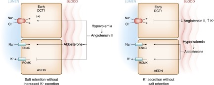
Potassium is the most abundant cation in the intracellular fluid, and maintaining the proper distribution of potassium across the cell membrane is critical for normal cell function. Long-term maintenance of potassium homeostasis is achieved by alterations in renal excretion of potassium in response to variations in intake. Understanding the mechanism and regulatory influences governing the internal distribution and renal clearance of potassium under normal circumstances can provide a framework for approaching disorders of potassium commonly encountered in clinical practice. This paper reviews key aspects of the normal regulation of potassium metabolism and is designed to serve as a readily accessible review for the well informed clinician as well as a resource for teaching trainees and medical students.

Keywords: aldosterone; collecting duct; potassium; renal physiology; tubular transport.


Figures

Figure 1.
Figure 1.
The cell model illustrates β2-adrenergic and insulin-mediated regulatory pathways for K+ uptake. β2-Adrenergic and insulin both lead to K+ uptake by stimulating the activity of the Na+-K+-ATPase pump primarily in skeletal muscle, but they do so through different signaling pathways. β2-Adrenergic stimulation leads to increased pump activity through a cAMP- and protein kinase A (PKA)–dependent pathway. Insulin binding to its receptor leads to phosphorylation of the insulin receptor substrate protein (IRS-1), which, in turn, binds to phosphatidylinositide 3-kinase (PI3-K). The IRS-1–PI3-K interaction leads to activation of 3-phosphoinositide–dependent protein kinase-1 (PDK1). The stimulatory effect of insulin on glucose uptake and K+ uptake diverge at this point. An Akt-dependent pathway is responsible for membrane insertion of the glucose transporter GLUT4, whereas activation of atypical protein kinase C (aPKC) leads to membrane insertion of the Na+-K+-ATPase pump (reviewed in ref. 3).
Figure 2.
Figure 2.
The effect of metabolic acidosis on internal K+ balance in skeletal muscle. (A) In metabolic acidosis caused by inorganic anions (mineral acidosis), the decrease in extracellular pH will decrease the rate of Na+-H+ exchange (NHE1) and inhibit the inward rate of Na+-3HCO3 cotransport (NBCe1 and NBCe2). The resultant fall in intracellular Na+ will reduce Na+-K+-ATPase activity, causing a net loss of cellular K+. In addition, the fall in extracellular HCO3 concentration will increase inward movement of Cl by Cl-HCO exchange, further enhancing K+ efflux by K+-Cl cotransport. (B) Loss of K+ from the cell is much smaller in magnitude in metabolic acidosis caused by an organic acidosis. In this setting, there is a strong inward flux of the organic anion and H+ through the monocarboxylate transporter (MCT; MCT1 and MCT4). Accumulation of the acid results in a larger fall in intracellular pH, thereby stimulating inward Na+ movement by way of Na+-H+ exchange and Na+-3HCO3 cotransport. Accumulation of intracellular Na+ maintains Na+-K+-ATPase activity, thereby minimizing any change in extracellular K+ concentration.
Figure 3.
Figure 3.
A cell model for K+ transport in the proximal tubule. K+ reabsorption in the proximal tubule primarily occurs through the paracellular pathway. Active Na+ reabsorption drives net fluid reabsorption across the proximal tubule, which in turn, drives K+ reabsorption through a solvent drag mechanism. As fluid flows down the proximal tubule, the luminal voltage shifts from slightly negative to slightly positive. The shift in transepithelial voltage provides an additional driving force favoring K+ diffusion through the low-resistance paracellular pathway. Experimental studies suggest that there may be a small component of transcellular K+ transport; however, the significance of this pathway is not known. K+ uptake through the Na+-K+-ATPase pump can exit the basolateral membrane through a conductive pathway or coupled to Cl. An apically located K+ channel functions to stabilize the cell negative potential, particularly in the setting of Na+-coupled cotransport of glucose and amino acids, which has a depolarizing effect on cell voltage.
Figure 4.
Figure 4.
A cell model for K+ transport in the thick ascending limb of Henle. K+ reabsorption occurs by both paracellular and transcellular mechanisms. The basolateral Na+-K+-ATPase pump maintains intracellular Na+ low, thus providing a favorable gradient to drive the apically located Na+-K+-2Cl cotransporter (an example of secondary active transport). The apically located renal outer medullary K+ (ROMK) channel provides a pathway for K+ to recycle from cell to lumen, and ensures an adequate supply of K+ to sustain Na+-K+-2Cl cotransport. This movement through ROMK creates a lumen-positive voltage, providing a driving force for passive K+ reabsorption through the paracellular pathway. Some of the K+ entering the cell through the cotransporter exits the cell across the basolateral membrane, accounting for transcellular K+ reabsorption. K+ can exit the cell through a conductive pathway or in cotransport with Cl. ClC-Kb is the primary pathway for Cl efflux across the basolateral membrane.
Figure 5.
Figure 5.
A cell model for K+ transport in the distal convoluted tubule (DCT). In the early DCT, luminal Na+ uptake is mediated by the apically located thiazide-sensitive Na+-Cl cotransporter. The transporter is energized by the basolateral Na+-K+-ATPase, which maintains intracellular Na+ concentration low, thus providing a favorable gradient for Na+ entry into the cell through secondary active transport. The cotransporter is abundantly expressed in the DCT1 but progressively declines along the DCT2. ROMK is expressed throughout the DCT and into the cortical collecting duct. Expression of the epithelial Na+ channel (ENaC), which mediates amiloride-sensitive Na+ absorption, begins in the DCT2 and is robustly expressed throughout the downstream connecting tubule and cortical collecting duct. The DCT2 is the beginning of the aldosterone-sensitive distal nephron (ASDN) as identified by the presence of both the mineralocorticoid receptor and the enzyme 11β-hydroxysteroid dehydrogenase II. This enzyme maintains the mineralocorticoid receptor free to only bind aldosterone by metabolizing cortisol to cortisone, the latter of which has no affinity for the receptor. Electrogenic-mediated K+ transport begins in the DCT2 with the combined presence of ROMK, ENaC, and aldosterone sensitivity. Electroneutral K+-Cl cotransport is present in the DCT and collecting duct. Conditions that cause a low luminal Cl concentration increase K+ secretion through this mechanism, which occurs with delivery of poorly reabsorbable anions, such as sulfate, phosphate, or bicarbonate.
Figure 6.
Figure 6.
The cell that is responsible for K+ secretion in the initial collecting duct and the cortical collecting duct is the principal cell. This cell possesses a basolateral Na+-K+-ATPase that is responsible for the active transport of K+ from the blood into the cell. The resultant high cell K+ concentration provides a favorable diffusion gradient for movement of K+ from the cell into the lumen. In addition to establishing a high intracellular K+ concentration, activity of this pump lowers intracellular Na+ concentration, thus maintaining a favorable diffusion gradient for movement of Na+ from the lumen into the cell. Both the movements of Na+ and K+ across the apical membrane occur through well defined Na+ and K+ channels.
Figure 7.
Figure 7.
Reabsorption of HCO3 in the distal nephron is mediated by apical H+ secretion by the α-intercalated cell. Two transporters secrete H+, a vacuolar H+-ATPase and an H+-K+-ATPase. The H+-K+-ATPase uses the energy derived from ATP hydrolysis to secrete H+ into the lumen and reabsorb K+ in an electroneutral fashion. The activity of the H+-K+-ATPase increases in K+ depletion and, thus, provides a mechanism by which K+ depletion enhances both collecting duct H+ secretion and K+ absorption.
Figure 8.
Figure 8.
Under normal circumstances, delivery of Na+ to the distal nephron is inversely associated with serum aldosterone levels. For this reason, renal K+ excretion is kept independent of changes in extracellular fluid volume. Hypokalemia caused by renal K+ wasting can be explained by pathophysiologic changes that lead to coupling of increased distal Na+ delivery and aldosterone or aldosterone-like effects. When approaching the hypokalemia caused by renal K+ wasting, one must determine whether the primary disorder is an increase in mineralocorticoid activity or an increase in distal Na+ delivery. EABV, effective arterial blood volume.
Figure 9.
Figure 9.
The aldosterone paradox refers to the ability of the kidney to stimulate NaCl retention with minimal K+ secretion under conditions of volume depletion and maximize K+ secretion without Na+ retention in hyperkalemia. With volume depletion (left panel), increased circulating angiotensin II (AII) levels stimulate the Na+-Cl cotransporter in the early DCT. In the ASDN, AII along with aldosterone stimulate the ENaC. In this latter segment, AII exerts an inhibitory effect on ROMK, thereby providing a mechanism to maximally conserve salt and minimize renal K+ secretion. When hyperkalemia or increased dietary K+ intake occurs with normovolemia (right panel), low circulating levels of AII or direct effects of K+ lead to inhibition of Na+-Cl cotransport activity along with increased activity of ROMK. As a result, Na+ delivery to the ENaC is optimized for the coupled electrogenic secretion of K+ through ROMK. As discussed in the text, with no lysine [K] 4 (WNK4) proteins are integrally involved in the signals by which the paradox is brought about. It should be emphasized the WNK proteins are part of a complex signaling network still being fully elucidated. The interested reader is referred to several recent reviews and advancements on this subject (,,–93).

References

    1. Palmer BF: A physiologic-based approach to the evaluation of a patient with hyperkalemia. Am J Kidney Dis 56: 387–393, 2010 - PubMed
    1. Foley K, Boguslavsky S, Klip A: Endocytosis, recycling, and regulated exocytosis of glucose transporter 4. Biochemistry 50: 3048–3061, 2011 - PubMed
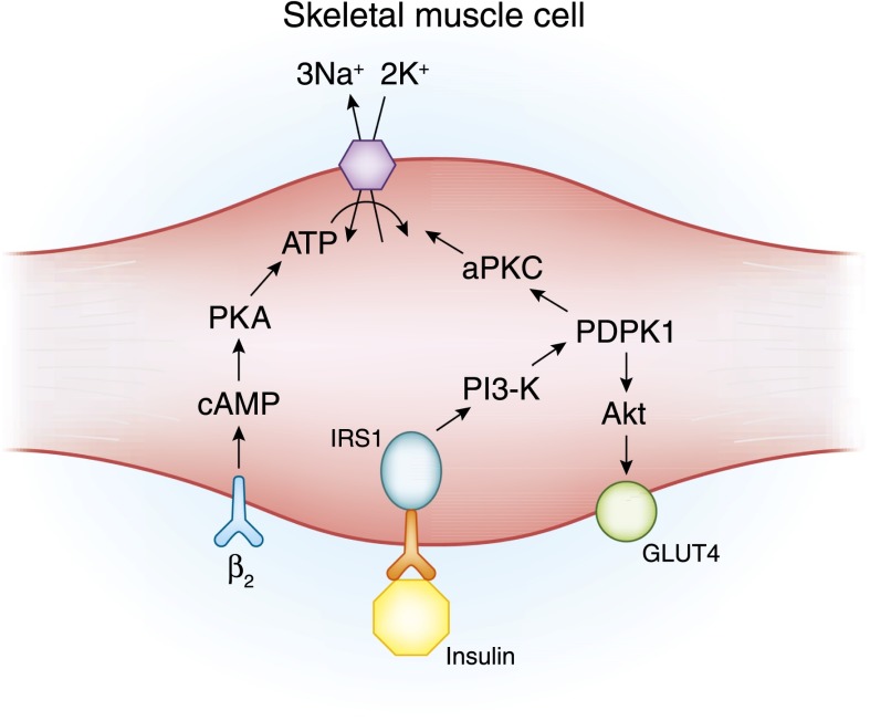
    1. Ho K: A critically swift response: Insulin-stimulated potassium and glucose transport in skeletal muscle. Clin J Am Soc Nephrol 6: 1513–1516, 2011 - PubMed
    1. Nguyen TQ, Maalouf NM, Sakhaee K, Moe OW: Comparison of insulin action on glucose versus potassium uptake in humans. Clin J Am Soc Nephrol 6: 1533–1539, 2011 - PMC - PubMed
    1. Alvestrand A, Wahren J, Smith D, DeFronzo RA: Insulin-mediated potassium uptake is normal in uremic and healthy subjects. Am J Physiol 246: E174–E180, 1984 - PubMed