. 2014 Apr 10;10(4):e1004275.
doi: 10.1371/journal.pgen.1004275. eCollection 2014 Apr.

Interplay of the serine/threonine-kinase StkP and the paralogs DivIVA and GpsB in pneumococcal cell elongation and division

Affiliations

Interplay of the serine/threonine-kinase StkP and the paralogs DivIVA and GpsB in pneumococcal cell elongation and division

Aurore Fleurie et al. PLoS Genet. .

Abstract

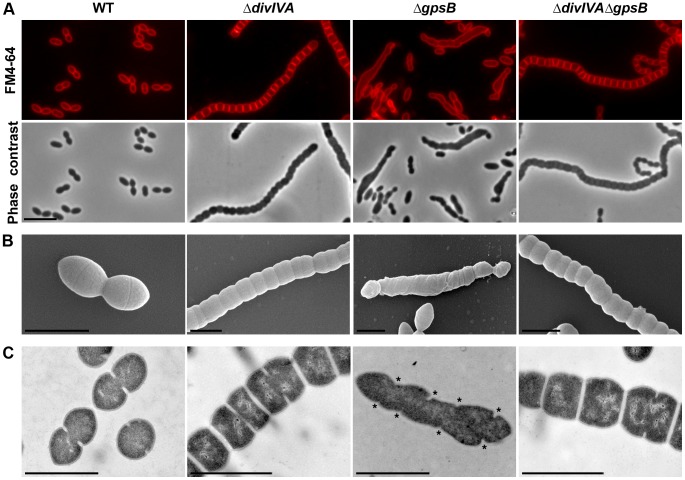
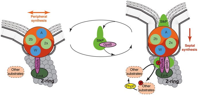
Despite years of intensive research, much remains to be discovered to understand the regulatory networks coordinating bacterial cell growth and division. The mechanisms by which Streptococcus pneumoniae achieves its characteristic ellipsoid-cell shape remain largely unknown. In this study, we analyzed the interplay of the cell division paralogs DivIVA and GpsB with the ser/thr kinase StkP. We observed that the deletion of divIVA hindered cell elongation and resulted in cell shortening and rounding. By contrast, the absence of GpsB resulted in hampered cell division and triggered cell elongation. Remarkably, ΔgpsB elongated cells exhibited a helical FtsZ pattern instead of a Z-ring, accompanied by helical patterns for DivIVA and peptidoglycan synthesis. Strikingly, divIVA deletion suppressed the elongated phenotype of ΔgpsB cells. These data suggest that DivIVA promotes cell elongation and that GpsB counteracts it. Analysis of protein-protein interactions revealed that GpsB and DivIVA do not interact with FtsZ but with the cell division protein EzrA, which itself interacts with FtsZ. In addition, GpsB interacts directly with DivIVA. These results are consistent with DivIVA and GpsB acting as a molecular switch to orchestrate peripheral and septal PG synthesis and connecting them with the Z-ring via EzrA. The cellular co-localization of the transpeptidases PBP2x and PBP2b as well as the lipid-flippases FtsW and RodA in ΔgpsB cells further suggest the existence of a single large PG assembly complex. Finally, we show that GpsB is required for septal localization and kinase activity of StkP, and therefore for StkP-dependent phosphorylation of DivIVA. Altogether, we propose that the StkP/DivIVA/GpsB triad finely tunes the two modes of peptidoglycan (peripheral and septal) synthesis responsible for the pneumococcal ellipsoid cell shape.


Conflict of interest statement

The authors have declared that no competing interests exist.

Figures

Figure 1
Figure 1. Morphology of WT, ΔdivIVA, ΔgpsB and ΔdivIVAΔgpsB cells.
(A) Phase contrast microscopy (lower panel) and FM4–64 membrane staining (upper panel) images of WT, ΔdivIVA, ΔgpsB and ΔdivIVAΔgpsB exponentially growing cells at 37°C in THY medium. Scale bar, 5 µm. (B) Scanning electron micrograph of WT, ΔdivIVA, ΔgpsB and ΔdivIVAΔgpsB cells. Scale bar, 1 µm. (C) Transmission electron micrograph of WT, ΔdivIVA, ΔgpsB and ΔdivIVAΔgpsB cells. Scale bar, 1 µm. Asterisks indicate defective septal initiations in staggered rows in the ΔgpsB cell.
Figure 2
Figure 2. Localization of PG synthesis in WT, ΔdivIVA, ΔgpsB and ΔdivIVAΔgpsB cells.
Phase contrast microscopy (left panel) and BADA labeling of PG (middle panel) images of WT, ΔdivIVA, ΔgpsB and ΔdivIVAΔgpsB exponentially growing cells pulsed with BADA 4 min each at 37°C in THY medium. Overlay between phase contrast (red) and BADA (green) labeling is shown. Arrows show helical organization of PG synthesis. Scale bar, 5 µm.
Figure 3
Figure 3. FtsZ localization in WT, ΔdivIVA, ΔgpsB and ΔdivIVAΔgpsB cells.
(A) FtsZ localization in WT, ΔdivIVA, ΔgpsB and ΔdivIVAΔgpsB cells. Phase contrast (left), GFP fluorescent signal (middle) and overlays (right) between phase contrast (red) and GFP (green) images are shown. Arrows show helical organization of FtsZ. Scale bar, 5 µm. See also the unprocessed image of FtsZ localization in ΔgpsB cells in Figure S5 showing that the FtsZ fluorescent signal is detected in all cells. (B) Immunofluorescence staining of fixed WT and ΔgpsB cells using anti-FtsZ polyclonal antibodies. DNA was counterstained with DAPI. Merged pictures show (upper panels) the overlay of FtsZ (red) and phase contrast images, and (lower panels) the overlay of FtsZ (red) and DAPI (blue). Higher magnifications of ΔgpsB cells highlighted with a white square are shown in the right row. Arrows show helical organization of FtsZ. Scale bar, 5 µm. (C) Fluorescence time-lapse microscopy of ΔgpsB cells producing FtsZ-GFP and grown in C+H medium at 30°C. Overlays between phase contrast (gray) and GFP (green) are shown. Stills are from Movie S2. Scale bar, 2 µm. Blue arrows point to cells in which the Z-ring helix-stretches until cell death. Red arrowheads point to helical structures of FtsZ. FtsZ-GFP is the only source of FtsZ in cells. ftsZ-gfp substitutes the native ftsZ gene at its chromosomal locus.
Figure 4
Figure 4. Localization of PBP2x, PBP2b, FtsW and RodA in WT, ΔdivIVA, ΔgpsB and ΔdivIVAΔgpsB cells.
Chromosomal copy of either pbp2x, pbp2b, ftsW or rodA were substituted for a gfp-fused gene in WT (A) or ΔgpsB (B), or ΔdivIVA (C), or ΔdivIVAΔgpsB (D) cells. Cells were grown in THY medium at 37°C. GFP (green) and phase-contrast (grey) images were taken from a typical field of exponentially growing cells. Merged pictures show the overlay of GFP fluorescence (green) and phase contrast images (red). Arrows show helical organization of GFP-PBP2x, GFP-PBP2b, FtsW-GFP and RodA-GFP. Scale bar, 5 µm. All fusion proteins are the only source of PBP2X, PBP2b, FtsW or RodA in the cells.
Figure 5
Figure 5. Localization of GFP fused PBP2x, PBP2b, FtsW or RodA together with FtsZ-RFP in WT and ΔgpsB cells.
Localization of FtsZ-RFP and either GFP-PBP2x, GFP-PBP2b, FtsW-GFP or RodA-GFP in WT (A) or ΔgpsB (B) cells grown at 37° in THY. Overlays between phase contrast (gray), GFP (green), and RFP (red) are shown on the right. Arrows show helical organization of FtsZ-RFP, GFP-PBP2x, GFP-PBP2b, FtsW-GFP and RodA-GFP. Scale bar, 5 µm. All fusion proteins are the only source of FtsZ, PBP2X, PBP2b, FtsW or RodA in cells. The fusion genes encoding these proteins substitute the corresponding native genes at their chromosomal locus.
Figure 6
Figure 6. Localization of DivIVA in WT and ΔgpsB cells.
(A) DivIVA-GFP localization in WT and ΔgpsB cells. Phase contrast (left), GFP fluorescent signal (middle) and overlays (right) between phase contrast (red) and GFP (green) images are shown. (B) Co-localization of FtsZ-RFP (red) and DivIVA-GFP (green) in WT and ΔgpsB cells. Overlays between phase contrast (gray), GFP (green), and RFP (red) are shown. Cells were grown to exponential phase in THY medium at 37°C. Arrows show helical organization of DivIVA-GFP and FtsZ-RFP. Scale bar, 5 µm. DivIVA-GFP and FtsZ-GFP are the only source of FtsZ and DivIVA in cells. ftsZ-gfp and divIVA-gfp substitute the native ftsZ and divIVA genes at their chromosomal locus, respectively.
Figure 7
Figure 7. Localization of GpsB and EzrA.
(A) Localization of GFP-GpsB in WT and ΔdivIVA cells. Expression of the gfp-gpsB fusion is under the control of the zinc-inducible Pzn promoter at the non-essential bgaA locus. (B) EzrA-GFP localization in WT, ΔgpsB, ΔdivIVA and ΔdivIVAΔgpsB cells. Phase contrast (left), GFP fluorescent signal (middle) and overlays (right) between phase contrast (red) and GFP (green) images are shown. Cells were grown to exponential phase in THY medium at 37°C. Arrows show helical organization of EzrA-GFP. Scale bar, 5 µm. EzrA-GFP is the only source of EzrA in cells. ezrA-gfp substitutes the native ezrA gene at its chromosomal locus.
Figure 8
Figure 8. Interplay of GpsB, DivIVA and StkP.
(A) Western immunoblot of whole-cell lysates from the wild type (WT), ΔstkP, gpsB::gfp-gpsB, ΔgpsB, ΔdivIVA and ΔgpsBΔdivIVA cells grown in THY at 37°C probed with anti-phosphothreonine antibodies. The same amounts (25 µg) of proteins were loaded in all gel lanes. Arrow indicates the signal observed around 15 kDa. The phosphorylation signal for DivIVA and StkP are indicated. (B) Western immunoblot of whole-cell lysates from wild type (WT) or gpsB::gfp-gpsB cells probed with anti-GFP antibodies. Purified GFP is used as control. Arrow indicates the signal observed for GFP-GpsB. (C) StkP localization using a GFP N-terminal fusion in WT, ΔgpsB, ΔdivIVA and ΔgpsBΔdivIVA cells. GFP (green) and phase-contrast (grey) images were taken from a typical field of exponentially grown cells in THY at 37°C. Merged pictures (lower panels) show the overlay of StkP (green) and phase contrast images (red). Scale bar, 5 µm. (D) Cell morphology of stkP-K42M cells deficient for DivIVA or GpsB expression. Cells producing a kinase dead-form of StkP (stkP-K42M, see [14]) were deleted either for divIVA or gpsB resulting thus in ΔdivIVA-stkP-K42M and ΔgpsB-stkP-K42M strains, respectively. Phase contrast microscopy (upper row) and FM4–64 membrane staining (lower row) images of ΔdivIVA-stkP-K42M (left panel) and ΔgpsB-stkP-K42M (right panel) exponentially growing cells at 37°C in THY medium. Scale bar, 5 µm.
Figure 9
Figure 9. Models for PG synthesis in S. pneumoniae.
In this model, a large membrane PG assembly complex (Yin Yang circle) contains both the septal (red) and the peripheral (orange) PG assembly machineries. The two transpeptidases PBP2x and PBP2b (noted 2x and 2b) and the two lipid-flippases FtsW and RodA (noted W and A) are indicated in green and blue, respectively. Non-phosphorylated forms of DivIVA and other StkP substrates are required for cell elongation and thus peripheral PG synthesis. GpsB is not per se involved in the production of the cross-wall, but is required at the septum to localize StkP (light green oval), to allow the phosphorylation of StkP substrates including DivIVA and to favor production of septal PG by down-regulating peripheral PG synthesis. The paralogs GpsB (pink oval) and DivIVA (purple oval) constitute a molecular switch that connects, together with EzrA (green oval), the Z-ring with the PG assembly complex. StkP kinase activity, counterbalanced by the phosphatase PhpP (yellow oval) and triggered by GpsB, modulates the function of a set of proteins (dashed ovals) including DivIVA . The StkP/DivIVA/GpsB triad is thus proposed to orchestrate and to finely tune production of septal and peripheral peptidoglycan synthesis responsible for the ovoid-shape of pneumococcus.
Figure 10
Figure 10. Alignment for GpsB and DivIVA proteins from several bacteria.
(A) Multiple sequence alignments of GpsB and DivIVA sequences from streptococci and Gram-positive bacteria. Protein sequences similar to that of pneumococcus GpsB and DivIVA were identified by BLAST searches and aligned using CLUSTALW. Spn: S. pneumoniae; Sag: S. agalactiae, Bsu: B. subtilis; Sta: S. aureus, Mtb: M. tuberculosis; Sco: S. coelicolor. Yellow highlights the potential coiled-coil motifs retrieved from UniProtKB/Swiss-Prot:Q8CWP9 and UniProtKB/Swiss-Prot:C1CIN3 entry annotations for Spn-DivIVA (residues 34–135 and 199–236) and Spn-GpsB (36–63) respectively. The PF05103 PFAM DivIVA family signatures are mapped as green open boxes for DivIVA and GpsB. When identified, phosphorylation sites are red boxed. The S. coelicolor DivIVA phosphopeptide containing unidentified phosphorylation sites are highlighted in orange letters. Identical residues are in pink letters and positions showing conservation of similar residues are in blue. Dots indicate gaps introduced in sequences during alignment computation. The figure was rendered with the ESPript server . (B) Multiple sequence alignments of GpsB sequences from streptococci. Protein sequences were aligned using CLUSTALW. Spy: S. pyogenes; Sag: S. agalactiae, Smu: S. mutans; Sth: S. thermophylus; Ssa: S. salivarius; Spn: S. pneumoniae; Smi: S. mitis, Sgo: S. gordonii. The PFAM PF05103 DivIVA family signatures are mapped as green boxes. Yellow highlights the potential coiled-coil motifs retrieved from UniProtKB/Swiss-Prot:C1CIN3 entry annotations for Spn-GpsB (36–63). The phosphothreonine identified for S. agalactiae GpsB is red boxed. Glutamic acids possibly mimicking threonine phosphorylation are black boxed with white letters. Identical residues are in pink letters and positions showing conservation of similar residues are in blue. Dots indicate gaps introduced in sequences during alignment computation. The figure was rendered with the ESPript server .

Similar articles

Cited by

References

    1. Young KD (2010) Bacterial shape: two-dimensional questions and possibilities. Annual review of microbiology 64: 223–240. - PMC - PubMed
    1. Egan AJ, Vollmer W (2012) The physiology of bacterial cell division. Annals of the New York Academy of Sciences 1277: 8–28. - PubMed
    1. Typas A, Banzhaf M, Gross CA, Vollmer W (2012) From the regulation of peptidoglycan synthesis to bacterial growth and morphology. Nature reviews Microbiology 10: 123–136. - PMC - PubMed
    1. Cabeen MT, Jacobs-Wagner C (2010) The bacterial cytoskeleton. Annual review of genetics 44: 365–392. - PubMed
    1. Flardh K (2010) Cell polarity and the control of apical growth in Streptomyces . Current opinion in microbiology 13: 758–765. - PubMed

Publication types

MeSH terms

LinkOut - more resources