

Molecular staging of lymph node-negative colon carcinomas by one-step nucleic acid amplification (OSNA) results in upstaging of a quarter of patients in a prospective, European, multicentre study
- PMID: 24722182
- PMCID: PMC4021519
- DOI: 10.1038/bjc.2014.170
Molecular staging of lymph node-negative colon carcinomas by one-step nucleic acid amplification (OSNA) results in upstaging of a quarter of patients in a prospective, European, multicentre study
Abstract
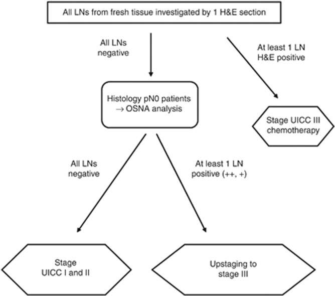
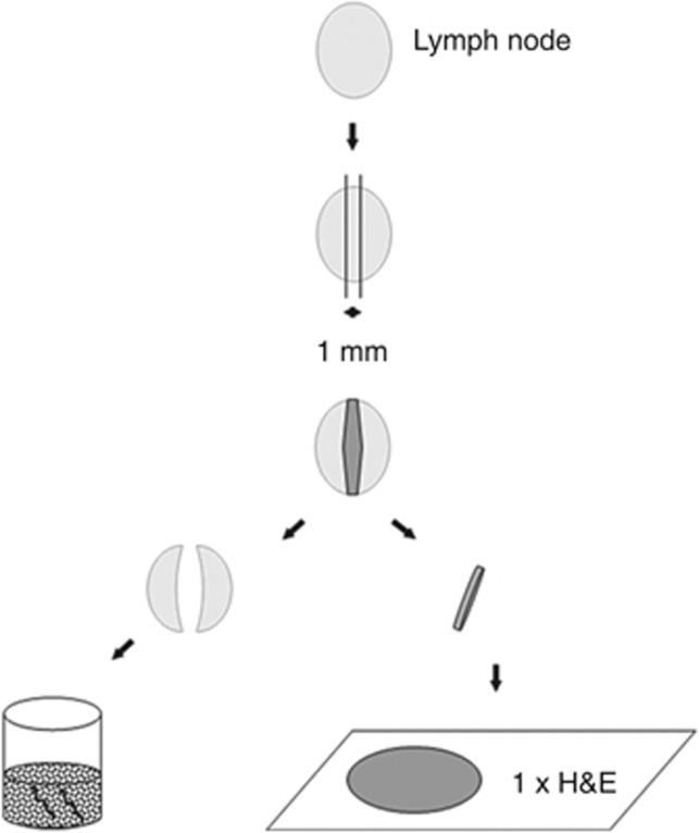
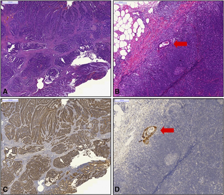
Background: Current histopathological staging procedures in colon carcinomas depend on midline division of the lymph nodes with one section of haematoxylin & eosin (H&E) staining only. By this method, tumour deposits outside this transection line may be missed and could lead to understaging of a high-risk group of stage UICC II cases, which recurs in ∼20% of cases. A new diagnostic semiautomated system, one-step nucleic acid amplification (OSNA), detects cytokeratin (CK) 19 mRNA in lymph node metastases and enables the investigation of the whole lymph node. The objective of this study was to assess whether histopathological pN0 patients can be upstaged to stage UICC III by OSNA.
Methods: Lymph nodes from patients who were classified as lymph node negative after standard histopathology (single (H&E) slice) were subjected to OSNA. A result revealing a CK19 mRNA copy number >250, which makes sure to detect mainly macrometastases and not isolated tumour cells (ITC) or micrometastases only, was regarded as positive for lymph node metastases based on previous threshold investigations.
Results: In total, 1594 pN0 lymph nodes from 103 colon carcinomas (median number of lymph nodes per patient: 14, range: 1-46) were analysed with OSNA. Out of 103 pN0 patients, 26 had OSNA-positive lymph nodes, resulting in an upstaging rate of 25.2%. Among these were 6/37 (16.2%) stage UICC I and 20/66 (30.3%) stage UICC II patients. Overall, 38 lymph nodes were OSNA positive: 19 patients had one, 3 had two, 3 had three, and 1 patient had four OSNA-positive lymph nodes.
Conclusions: OSNA resulted in an upstaging of over 25% of initially histopathologically lymph node-negative patients. OSNA is a standardised, observer-independent technique, allowing the analysis of the whole lymph node. Therefore, sampling bias due to missing investigation of certain lymph node tissue can be avoided, which may lead to a more accurate staging.
Figures
References
-
- Armitage P. Trends for linear trends in proportions and frequencies. Biometrics. 1955;11:375–386.
-
- Baloch ZW, Abraham S, Roberts S, LiVolsi VA. Differential expression of cytokeratins in follicular variant of papillary carcinoma: an immunohistochemical study and its diagnostic utility. Hum Pathol. 1999;30:1166–1171. - PubMed
-
- Croner RS, Fortsch T, Bruckl WM, Rodel F, Rodel C, Papadopoulos T, Brabletz T, Kirchner T, Sachs M, Behrens J, Klein-Hitpass L, Sturzl M, Hohenberger W, Lausen B. Molecular signature for lymphatic metastasis in colorectal carcinomas. Ann Surg. 2008;247:803–810. - PubMed
-
- Croner RS, Merkel S, Papadopoulos T, Schellerer V, Hohenberger W, Goehl J. Multivisceral resection for colon carcinoma. Dis Colon Rectum. 2009;52:1381–1386. - PubMed
Publication types
MeSH terms
Substances
LinkOut - more resources
Full Text Sources
Other Literature Sources
Research Materials
