. 2014 Apr 10;10(4):e1004287.
doi: 10.1371/journal.pgen.1004287. eCollection 2014 Apr.

Unexpected role of the steroid-deficiency protein ecdysoneless in pre-mRNA splicing

Affiliations

Unexpected role of the steroid-deficiency protein ecdysoneless in pre-mRNA splicing

Ann-Katrin Claudius et al. PLoS Genet. .

Abstract

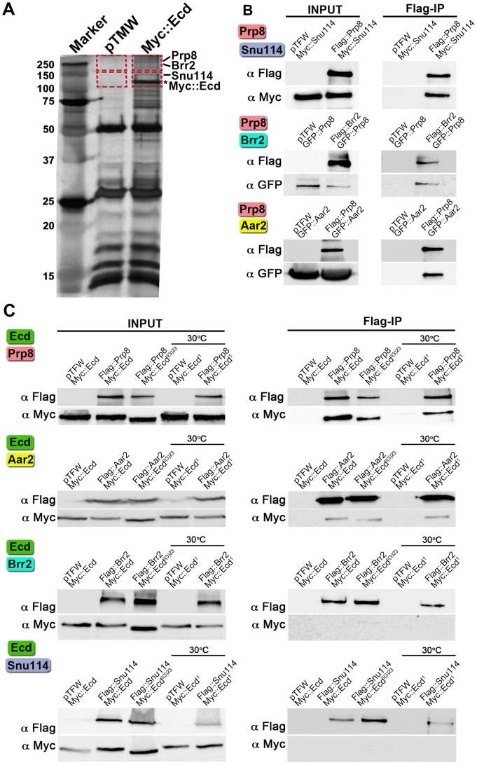
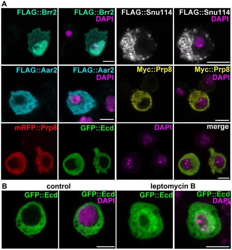
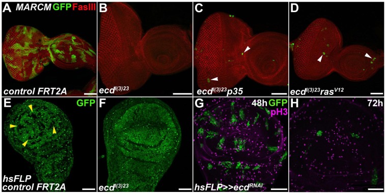
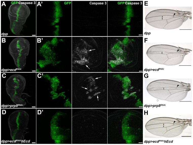
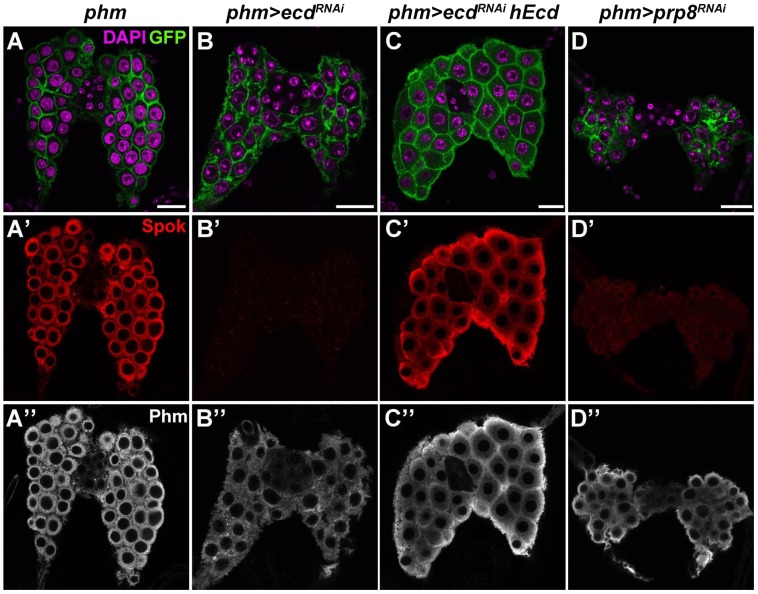
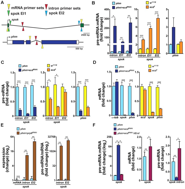
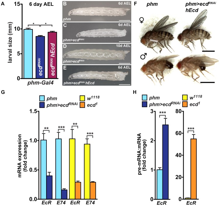
The steroid hormone ecdysone coordinates insect growth and development, directing the major postembryonic transition of forms, metamorphosis. The steroid-deficient ecdysoneless1 (ecd1) strain of Drosophila melanogaster has long served to assess the impact of ecdysone on gene regulation, morphogenesis, or reproduction. However, ecd also exerts cell-autonomous effects independently of the hormone, and mammalian Ecd homologs have been implicated in cell cycle regulation and cancer. Why the Drosophila ecd1 mutants lack ecdysone has not been resolved. Here, we show that in Drosophila cells, Ecd directly interacts with core components of the U5 snRNP spliceosomal complex, including the conserved Prp8 protein. In accord with a function in pre-mRNA splicing, Ecd and Prp8 are cell-autonomously required for survival of proliferating cells within the larval imaginal discs. In the steroidogenic prothoracic gland, loss of Ecd or Prp8 prevents splicing of a large intron from CYP307A2/spookier (spok) pre-mRNA, thus eliminating this essential ecdysone-biosynthetic enzyme and blocking the entry to metamorphosis. Human Ecd (hEcd) can substitute for its missing fly ortholog. When expressed in the Ecd-deficient prothoracic gland, hEcd re-establishes spok pre-mRNA splicing and protein expression, restoring ecdysone synthesis and normal development. Our work identifies Ecd as a novel pre-mRNA splicing factor whose function has been conserved in its human counterpart. Whether the role of mammalian Ecd in cancer involves pre-mRNA splicing remains to be discovered.


Conflict of interest statement

The authors have declared that no competing interests exist.

Figures

Figure 1
Figure 1. Interactions of Ecd with proteins of the U5 snRNP complex.
(A) Extracts from Drosophila S2 cells expressing the Myc::Ecd fusion protein or the Myc tag alone (pTMW) were affinity purified using the Myc epitope and resolved on silver-stained SDS-PAGE. Mass spectrometry on the gel sections (indicated by rectangles) identified Prp8, Brr2, and Snu114 proteins associated with Myc::Ecd (asterisk). (B) Drosophila Prp8 interacts with orthologs of known spliceosomal proteins: Snu114, Brr2, and Aar2. (C) Flag-tagged Prp8 and Aar2 but not Brr2 and Snu114 proteins bound Ecd or its mutant variants as assessed by Flag immunoprecipitation (Flag-IP). Cells expressing the temperature-sensitive Ecd1 mutant were upshifted to 30°C 45 min prior to lysis. In all assays (B, C), the pairs of proteins were expressed in S2 cells and their epitope tags were used for Flag-IP and immunoblot detection as indicated. The empty pTFW vector expressing the Flag peptide alone was included in control assays.
Figure 2
Figure 2. Subcellular localization of Ecd and the U5 snRNP proteins.
(A) Brr2 was enriched in cell nuclei while Ecd, Prp8, Snu114, and Aar2 were detected in the cytoplasm. The Flag and Myc epitope-tagged proteins were expressed in S2 cells; mRFP::Prp8 and GFP::Ecd (bottom row) were co-transfected. (B) Inhibition of nuclear export with leptomycin B resulted in nuclear retention of the GFP::Ecd protein. Nuclei are stained with DAPI. All images are single confocal sections. Scale bars, 5 µm.
Figure 3
Figure 3. Ecd is required for survival of imaginal disc cells.
(A–F) In contrast to abundant, sizable control clones (green in A and GFP-negative in E, yellow arrowheads), ecdl(3)23 homozygous mutant clones could be recovered neither in eye/antennal (B) nor wing (F) imaginal discs. Simultaneous inhibition of apoptosis with a pan-caspase inhibitor p35 (ecdl(3) 23 p35) (C) or cell growth enhancement with activated RasV12 (ecdl(3) 23 rasV12) (D) yielded rare and extremely small clones in eye/antennal discs (white arrowheads). Fasciclin III (FasIII) staining of lateral cell membranes reveals the overall tissue morphology. (G–H) ecdRNAi clones (green) were eliminated from the wing imaginal epithelium within 72 h after induction using the heat-shock flip-out technique. Only rare ecdRNAi cells labeled phospho-histone 3 (pH3) positive relative to many mitotically active surrounding control cells. Scale bars, 50 µm.
Figure 4
Figure 4. Depletion of Ecd or Prp8 causes apoptotic phenotypes in developing wings.
(A–D) RNAi knockdown of ecd (B) and prp8 (C) induced with the dpp-Gal4 driver disrupted the regular pattern of the dpp expression domain (visualized by co-expression of GFP) in third-instar wing imaginal discs and caused appearance of apoptotic, Caspase 3 positive cells (B′, C′, white arrows). Expression of human Ecd (hEcd) averted the apoptotic phenotype caused by ecd RNAi (D, D′). (E–H) The intervein region corresponding to the dpp expression domain was reduced (double arrows) and the anterior crossvein (arrowhead) was lost in the wings of dpp>ecdRNAi (F) and dpp>prp8RNAi (G) adult flies. The ecd RNAi phenotype was rescued by expression of hEcd (H). Images in (A–D) are maximum-intensity projections of multiple confocal sections; panels A′ through D′ are higher-magnification, single sections from the corresponding images in (A–D). Scale bars are 50 µm (A–D), 20 µm (A′–D′), and 1 mm (E–H).
Figure 5
Figure 5. Ecd and Prp8 are required for expression of Spok in the PG.
(A–D) Relative to control PG dissected 6 days AEL (A′, A″), expression of the Spok protein (B′) was undetected while the Phm signal (B″) was weakened in the PG of phm>ecdRNAi (B) and phm>prp8RNAi (D′, D″) larvae. Note the moderate reduction in size of PG cells and nuclei in phm>ecdRNAi (B) compared to a more severe PG deterioration in phm>prp8RNAi larvae (D). Expression of hEcd restored Spok and Phm expression (C′, C″) and improved the morphology of the Ecd-deficient PG (C). Cell membranes are decorated with CD8::GFP; DAPI stains the nuclei. Panels show single confocal sections. Scale bars, 20 µm. See Figure S1 for RNAi-mediated depletion of Ecd.
Figure 6
Figure 6. Loss of Ecd interferes with splicing of spok pre-mRNA.
(A) Schematic of Drosophila spok and phm gene loci. Open and colored boxes represent untranslated and translated exons, respectively; black arrows point in the direction of transcription. Colored arrowheads mark the positions of primer pairs used to discriminate between pre-mRNA and mRNA species by qRT-PCR: yellow, in exons separated by an intron; red, within an intron; green and blue, spanning exon-intron boundaries of the first (EI1) and second (EI2) spok exon, respectively. (B) Pre-mRNA:mRNA ratios, determined by qRT-PCR with primers depicted in (A), were significantly elevated for spok but not for phm in third-instar phm>ecdRNAi larvae and in ecd1 mutants at 29°C. (C, D) spok and phm pre-mRNA (C) and mRNA (D) levels decreased in phm>ecdRNAi and ecd1 larvae. Levels of α-tubulin 84B mRNA did not change upon ecd RNAi and expression of ecd itself was unaffected by the ecd1 mutation (D). (E) RNAi knockdown of prp8 in the PG diminished spok mRNA, whereas unspliced transcripts accumulated, causing the pre-mRNA:mRNA ratio to rise dramatically. (F) Expression of hEcd in the Ecd-deficient PG restored normal spok transcription and pre-mRNA splicing as judged from restored levels of pre-mRNA, mRNA, and their ratio. In all experiments, qRT-PCR was performed with total RNA from whole larvae 6 days AEL, and levels of rp49 transcripts were used for normalization. The pre-mRNA:mRNA ratios in (B, E, F) were calculated from the normalized qRT-PCR data by dividing values obtained with intron primer sets (A, red triangles) or primers spanning exon-intron boundaries (green or blue triangles) with values obtained using mRNA-specific primers (yellow triangles). Data are mean ± S.E.M; n≥4; *p<0.05, **p<0.01, and ***p<0.001.
Figure 7
Figure 7. PG-specific loss of Ecd results in systemic effects.
(A–E) Growth of phm>ecdRNAi larvae was retarded. Although reaching the third instar, phm>ecdRNAi larvae were smaller on day 6 AEL (A, C) compared to controls (B) or larvae supplemented with hEcd (phm>ecdRNAi hEcd) (E). Deficiency of Ecd in the PG prevented metamorphosis, generating permanent, eventually lethal larvae (D). (F) Expression of hEcd in the Ecd-deficient PG restored development of normally sized adults. (G) Expression of EcR and E74 (as assessed by qRT-PCR with primer sets detecting all alternatively spliced mRNA isoforms of each gene, see Table S1) was lowered to a similar degree in phm>ecdRNAi and in ecd1 larvae at 29°C. (H) The EcR pre-mRNA:mRNA ratio increased much more dramatically in ecd1 mutants at 29°C than in phm>ecdRNAi larvae. Data are mean ± S.E.M; n≥4; **p<0.01, and ***p<0.001.

References

    1. Thummel CS (2001) Molecular mechanisms of developmental timing in C. elegans and Drosophila. Dev Cell 1: 453–465. - PubMed
    1. Mirth CK, Riddiford LM (2007) Size assessment and growth control: how adult size is determined in insects. Bioessays 29: 344–355. - PubMed
    1. Yamanaka N, Rewitz KF, O'Connor MB (2013) Ecdysone control of developmental transitions: lessons from Drosophila research. Annu Rev Entomol 58: 497–516. - PMC - PubMed
    1. Ou Q, King-Jones K (2013) What goes up must come down: transcription factors have their say in making ecdysone pulses. Curr Top Dev Biol 103: 35–71. - PubMed
    1. Rewitz KF, Yamanaka N, O'Connor MB (2013) Developmental checkpoints and feedback circuits time insect maturation. Curr Top Dev Biol 103: 1–33. - PMC - PubMed

Publication types