

Transition into driven equilibrium of the balanced steady-state free precession as an ultrafast multisection T2-weighted imaging of the brain
- PMID: 24722304
- PMCID: PMC7965149
- DOI: 10.3174/ajnr.A3863
Transition into driven equilibrium of the balanced steady-state free precession as an ultrafast multisection T2-weighted imaging of the brain
Abstract
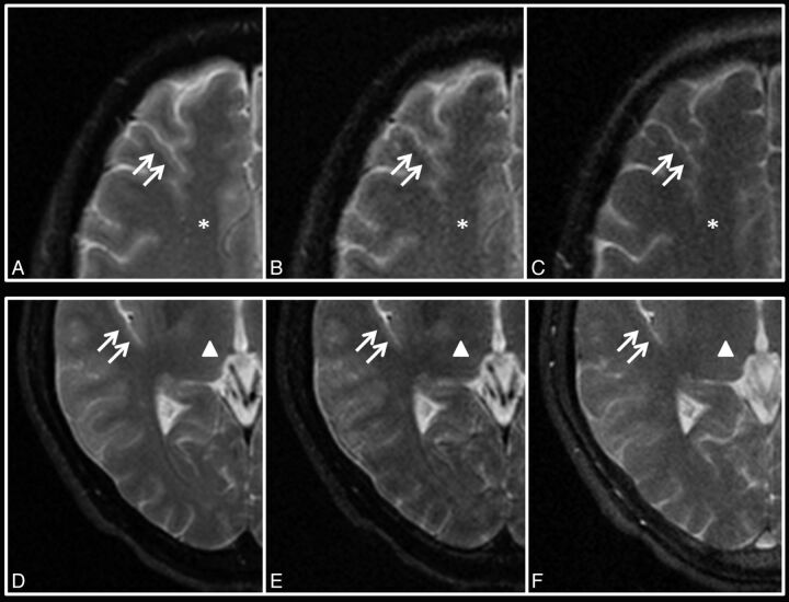
Background and purpose: Current T2-weighted imaging takes >3 minutes to perform, for which the ultrafast transition into driven equilibrium (TIDE) technique may be potentially helpful. This study qualitatively and quantitatively evaluates the imaging of transition into driven equilibrium of the balanced steady-state free precession (TIDE) compared with TSE and turbo gradient spin-echo on T2-weighted MR images.
Materials and methods: Thirty healthy volunteers were examined with T2-weighted images by using TIDE, TSE, and turbo gradient spin-echo sequences. Imaging was evaluated qualitatively by 2 independent observers on the basis of a 4-point rating scale regarding contrast characteristics and artifacts behavior. Image SNR and contrast-to-noise ratio were quantitatively assessed.
Results: TIDE provided T2-weighted contrast similar to that in TSE and turbo gradient spin-echo with only one-eighth of the scan time. TIDE showed gray-white matter differentiation and iron-load sensitivity inferior that of TSE and turbo gradient spin-echo, but with improved motion artifacts reduction on qualitative scores. Nonmotion ghosting artifacts were uniquely found in TIDE images. The overall SNRs of TSE were 1.9-2.0 times those of turbo gradient spin-echo and 1.7-2.2 times of those of TIDE for brain tissue (P < .0001). TIDE had a higher contrast-to-noise ratio than TSE (P = .169) and turbo gradient spin-echo (P < .0001) regarding non-iron-containing gray matter versus white matter. TIDE had a lower contrast-to-noise ratio than turbo gradient spin-echo and TSE (P < .0001) between iron-containing gray matter and white matter.
Conclusions: TIDE provides T2-weighted images with reduced scan times and reduced motion artifacts compared with TSE and turbo gradient spin-echo with the trade-off of reduced SNR and poorer gray-white matter differentiation.
© 2014 by American Journal of Neuroradiology.
Figures


Similar articles
-
T2-weighted MR imaging of the liver: qualitative and quantitative comparison of SPACE MR imaging with turbo spin-echo MR imaging.Eur J Radiol. 2013 Nov;82(11):e655-61. doi: 10.1016/j.ejrad.2013.07.020. Epub 2013 Aug 17. Eur J Radiol. 2013. PMID: 23957939
-
7T MRI-Histologic Correlation Study of Low Specific Absorption Rate T2-Weighted GRASE Sequences in the Detection of White Matter Involvement in Multiple Sclerosis.J Neuroimaging. 2015 May-Jun;25(3):370-8. doi: 10.1111/jon.12238. Epub 2015 Apr 21. J Neuroimaging. 2015. PMID: 25898858 Free PMC article.
-
Intrinsic fat suppression in TIDE balanced steady-state free precession imaging.Magn Reson Med. 2006 Dec;56(6):1328-35. doi: 10.1002/mrm.21084. Magn Reson Med. 2006. PMID: 17089365
-
[PSIF/CE-FAST (fast gradient echo) and TSE (turbo spin echo)].Nihon Rinsho. 1998 Nov;56(11):2773-7. Nihon Rinsho. 1998. PMID: 9847597 Review. Japanese.
-
Cardiovascular magnetic resonance physics for clinicians: part I.J Cardiovasc Magn Reson. 2010 Nov 30;12(1):71. doi: 10.1186/1532-429X-12-71. J Cardiovasc Magn Reson. 2010. PMID: 21118531 Free PMC article. Review.
References
-
- Juan CJ, Chen CY, Liu YJ, et al. . Acute putaminal necrosis and white matter demyelination in a child with subnormal copper metabolism in Wilson disease: MR imaging and spectroscopic findings. Neuroradiology 2005;47:401–05 - PubMed
-
- Forsting M. MR imaging of the brain: metabolic and toxic white matter diseases. Eur Radiol 1999;9:1061–65 - PubMed
-
- Patel MR, Klufas RA, Shapiro AW. MR imaging of diseases of the brain: comparison of GRASE and conventional spin-echo T2-weighted pulse sequences. AJR Am J Roentgenol 1995;165:963–66 - PubMed
-
- Hennig J, Nauerth A, Friedburg H. RARE imaging: a fast imaging method for clinical MR. Magn Reson Med 1986;3:823–33 - PubMed
Publication types
MeSH terms
LinkOut - more resources
Full Text Sources
Other Literature Sources
Medical