. 2014 Nov;25(11):2573-83.
doi: 10.1681/ASN.2013050565. Epub 2014 Apr 10.

Renal-retinal ciliopathy gene Sdccag8 regulates DNA damage response signaling

Affiliations

Renal-retinal ciliopathy gene Sdccag8 regulates DNA damage response signaling

Rannar Airik et al. J Am Soc Nephrol. 2014 Nov.

Abstract

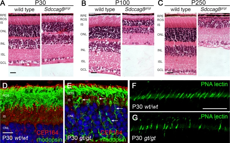
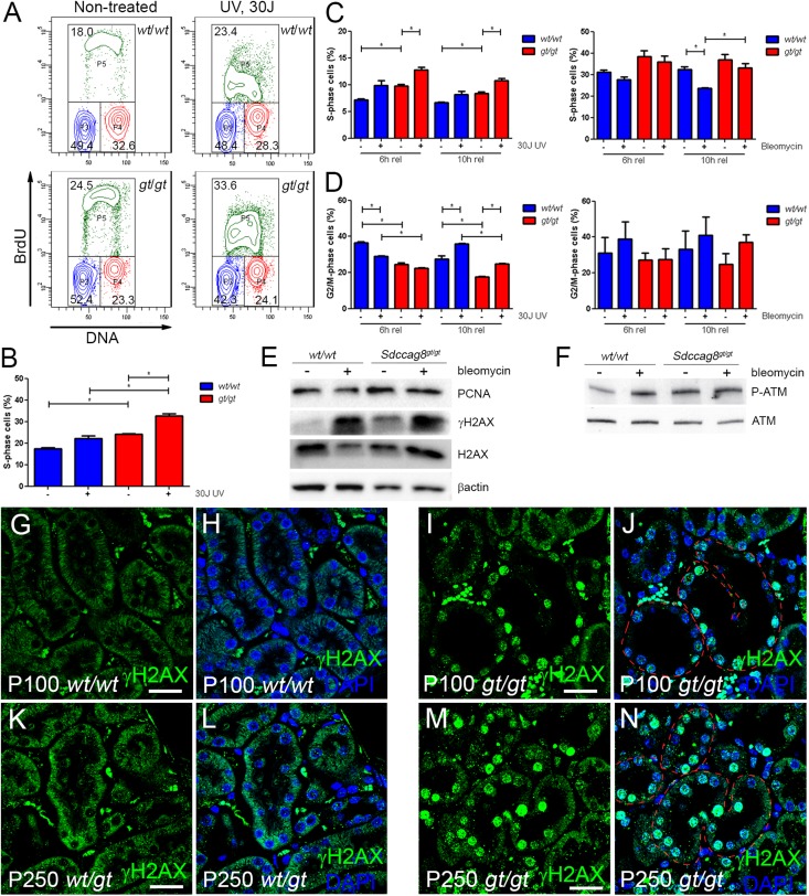
Nephronophthisis-related ciliopathies (NPHP-RCs) are developmental and degenerative kidney diseases that are frequently associated with extrarenal pathologies such as retinal degeneration, obesity, and intellectual disability. We recently identified mutations in a gene encoding the centrosomal protein SDCCAG8 as causing NPHP type 10 in humans. To study the role of Sdccag8 in disease pathogenesis, we generated a Sdccag8 gene-trap mouse line. Homozygous Sdccag8(gt/gt) mice lacked the wild-type Sdccag8 transcript and protein, and recapitulated the human phenotypes of NPHP and retinal degeneration. These mice exhibited early onset retinal degeneration that was associated with rhodopsin mislocalization in the photoreceptors and reduced cone cell numbers, and led to progressive loss of vision. By contrast, renal histologic changes occurred later, and no global ciliary defects were observed in the kidneys. Instead, renal pathology was associated with elevated levels of DNA damage response signaling activity. Cell culture studies confirmed the aberrant activation of DNA damage response in Sdccag8(gt/gt)-derived cells, characterized by elevated levels of γH2AX and phosphorylated ATM and cell cycle profile abnormalities. Our analysis of Sdccag8(gt/gt) mice indicates that the pleiotropic phenotypes in these mice may arise through multiple tissue-specific disease mechanisms.


Figures

Figure 1.
Figure 1.
Sdccag8 is expressed in kidney and lung epithelia. β-Galactosidase activity staining in the urogenital system at E16.5 demonstrates Sdccag8 expression in the corticomedullary region (arrows) and in the CCDs (arrowheads) in Sdccag8wt/gt kidneys (A and B). (C and D) Sdccag8 expression is maintained in the collecting ducts of the kidneys in two-week-old (C) and adult (D) mice. (E) β-Galactosidase activity staining on E16.5 whole lung demonstrates Sdccag8 expression in the bronchi and bronchioles. (F) Analysis of a cross-section through the X-gal–stained lung at higher resolution confirms Sdccag8 expression localization to the epithelial layer of the bronchioles and its absence from alveoli (delineated with a dashed line) and surrounding stromal mesenchyme. Notice the presence of lacZ-negative cells (white arrows) in the bronchioles. (G–I) CETN2-GFP expression in the bronchiole epithelium (G) overlaps (I) with SDCCAG8 antibody staining (H) in the E16.5 mouse lung section, indicating that Sdccag8-positive cells correspond to the progenitors of the multiciliated cells in the lung. Bar, 1 mm in A and B; 400 μm in C; 2 mm in D and E; 200 μm in F; 25 μm in G–I.
Figure 2.
Figure 2.
Sdccag8gt/gt mice develop nephronophthisis. (A–C) H&E staining of sagittal sections of P100 kidneys shows the formation of cortical cysts in Sdccag8gt/gt kidneys (B and C). (D and E) Analysis of H&E staining on sagittal sections of P250 kidneys shows loss of corticomedullary differentiation and progressive cyst formation in Sdccag8gt/gt kidneys (E). (F–H) H&E-stained Sdccag8wt/wt (F) and Sdccag8gt/gt (G and H) kidney sections demonstrate the variation in tubular cyst size and amount of interstitial infiltrate in Sdccag8gt/gt kidneys at P100 (G and H). (I–K) By P250, kidney cysts and interstitial infiltrate have replaced most of the renal parenchyma in Sdccag8gt/gt kidneys (J and K). Glomerular cysts are marked with asterisks. (L) The kidney weight to body weight ratio is not changed in Sdccag8gt/gt mice at P100 (mean wt/wt 1.667±0.09516, gt/gt 1.732±0.09247); however, the ratio is significantly increased in P250 Sdccag8gt/gt mice (mean wt/wt 1.554±0.07007, gt/gt 2.151±0.08878; *P<0.05). (M) The kidney cyst index is low in P100 Sdccag8gt/gt mice (mean 4.270±2.024), but is greatly increased and with noticeable variation in P250 mice (mean 21.91±8.662; *P<0.05). H&E, hematoxylin and eosin; wt, Sdccag8 wild-type allele; gt, Sdccag8 gene-trap allele; KW/BW, kidney weight to body weight ratio. Bar, 2 mm in A–E; 100 μm in F–K.
Figure 3.
Figure 3.
Sdccag8gt/gt mice develop fibrosis. (A–D) Masson trichrome staining reveals no collagen deposits in Sdccag8gt/gt kidneys at P100 (B); however, extensive fibrosis is visible in P250 Sdccag8gt/gt kidneys (D). (E) The fibrosis index is significantly increased in Sdccag8gt/gt mice between P100 (mean 10.50±10.50) and P250 (mean 47.00±12.41; *P<0.05). (F and G) LTA-FITC and DBA-rhodamine staining on kidney sections demonstrates the formation of cysts in the CCDs (DBA) and not in the proximal tubules (LTA) of Sdccag8gt/gt kidneys at P100. (H and I) Immunofluorescence staining with α-SMA-rhodamine and LTA-FITC demonstrate the presence of myofibroblasts in the proximity of a nascent cyst (I, white arrow). (J and K) Immunofluorescence staining with αSMA-FITC and DCT marker TSC show that besides CCDs, cysts also originate from DCTs (marked with white asterisks) in P100 Sdccag8gt/gt kidneys (K). αSMA-FITC staining shows the myofibroblasts surrounding the affected tubules. wt, Sdccag8 wild-type allele; gt, Sdccag8 gene-trap allele; α-SMA, α-smooth muscle actin. Bar, 100 μm in A–D; 100 μm in F–K.
Figure 4.
Figure 4.
Sdccag8 is required for photoreceptor maintenance. (A–C) Histologic analysis of H&E-stained wild-type and Sdccag8gt/gt retinas at P30 (A), P100 (B), and P250 (C) show progressive degeneration of photoreceptor inner segments and outer segments, as well as severe reduction of the outer nuclear layer in Sdccag8gt/gt mice. (D and E) Immunolocalization of CEP164 and rhodopsin in the P30 retina shows rhodopsin accumulation (white arrows) at the photoreceptor inner segment plasma membrane and cell bodies in Sdccag8gt/gt retina (E). (F and G) PNA-FITC lectin staining of cone sheaths at P30 demonstrates a reduction in cone cell number in Sdccag8gt/gt retina (G). H&E, hematoxylin and eosin; RPE, retinal pigment epithelium; ROS, retinal outer segment; IS, inner segment; ONL, outer nuclear layer; INL, inner nuclear layer; ISL, inner plexiform layer; GCL, ganglion cell layer; wt, Sdccag8 wild-type allele; gt, Sdccag8 gene-trap allele; PNA, peanut agglutinin lectin. Bar, 100 μm in A–C; 10 μm in D and E; 50 μm in F and G.
Figure 5.
Figure 5.
Sdccag8 inactivation causes S-phase delay and replication stress. (A and B) The cell cycle profile of Immorto;Sdccag8wt/wt and Immorto;Sdccag8gt/gt nonsynchronized cells demonstrates significant accumulation of Sdccag8gt/gt cells in the S phase (P<0.05; n=3) compared with the wild-type control (P<0.05, n=3). The number of cells in the S phase is further increased after treatment with 30 J UV light. (C) Sdccag8gt/gt cells synchronized with double thymidine block show a delay in S-phase progression, which is further increased after treatment with UV light. (D) Sdccag8gt/gt cells are competent in G2/M check point activation, both in response to UV light and bleomycin treatment. (E) Sdccag8gt/gt cells have increased PCNA levels, reflecting their extended duration in the S phase. PCNA levels do not change in response to bleomycin treatment, which is in agreement with the cell cycle analysis data. Increased γH2AX levels in cultured Sdccag8gt/gt cells are a sign of replication stress. H2AX and β-actin are used as loading controls. (F) Phosphorylated ATM levels are low in cultured wild-type cells, which become elevated upon bleomycin treatment. By contrast, Sdccag8gt/gt cells display high phosphorylated ATM levels even without treatment. ATM is used as a loading control. (G–J) Immunofluorescence staining with anti-γH2AX antibody on P100 Sdccag8wt/wt and Sdccag8gt/gt kidneys shows no γH2AX staining in control kidneys (G and H), whereas the dilated tubules (red dashed lines) in Sdccag8gt/gt kidneys are positive for γH2AX (I and J). (K–N) P250 Sdccag8wt/gt kidneys are negative for gH2AX staining (K and L), while Sdccag8gt/gt kidneys show positive staining in dilated and nondilated nephron epithelium (red dashed lines) and fibrotic mesenchyme (M and N). wt, Sdccag8 wild-type allele; gt, Sdccag8 gene-trap allele; PCNA, proliferating cell nuclear antigen. Bar, 25 μm in G–J; 20 μm in K–N.

References

    1. Hildebrandt F, Benzing T, Katsanis N: Ciliopathies. N Engl J Med 364: 1533–1543, 2011 - PMC - PubMed
    1. Hurd TW, Hildebrandt F: Mechanisms of nephronophthisis and related ciliopathies. Nephron, Exp Nephrol 118: e9–e14, 2011 - PMC - PubMed
    1. Otto EA, Hurd TW, Airik R, Chaki M, Zhou W, Stoetzel C, Patil SB, Levy S, Ghosh AK, Murga-Zamalloa CA, van Reeuwijk J, Letteboer SJ, Sang L, Giles RH, Liu Q, Coene KL, Estrada-Cuzcano A, Collin RW, McLaughlin HM, Held S, Kasanuki JM, Ramaswami G, Conte J, Lopez I, Washburn J, Macdonald J, Hu J, Yamashita Y, Maher ER, Guay-Woodford LM, Neumann HP, Obermüller N, Koenekoop RK, Bergmann C, Bei X, Lewis RA, Katsanis N, Lopes V, Williams DS, Lyons RH, Dang CV, Brito DA, Dias MB, Zhang X, Cavalcoli JD, Nürnberg G, Nürnberg P, Pierce EA, Jackson PK, Antignac C, Saunier S, Roepman R, Dollfus H, Khanna H, Hildebrandt F: Candidate exome capture identifies mutation of SDCCAG8 as the cause of a retinal-renal ciliopathy. Nat Genet 42: 840–850, 2010 - PMC - PubMed
    1. Schaefer E, Zaloszyc A, Lauer J, Durand M, Stutzmann F, Perdomo-Trujillo Y, Redin C, Bennouna Greene V, Toutain A, Perrin L, Gérard M, Caillard S, Bei X, Lewis RA, Christmann D, Letsch J, Kribs M, Mutter C, Muller J, Stoetzel C, Fischbach M, Marion V, Katsanis N, Dollfus H: Mutations in SDCCAG8/NPHP10 cause Bardet-Biedl syndrome and are associated with penetrant renal disease and absent polydactyly. Mol Syndromol 1: 273–281, 2011 - PMC - PubMed
    1. Kenedy AA, Cohen KJ, Loveys DA, Kato GJ, Dang CV: Identification and characterization of the novel centrosome-associated protein CCCAP. Gene 303: 35–46, 2003 - PubMed

Publication types

MeSH terms