. 2014 Nov;25(11):2511-25.
doi: 10.1681/ASN.2013050522. Epub 2014 Apr 10.

Alterations in the ubiquitin proteasome system in persistent but not reversible proteinuric diseases

Affiliations

Alterations in the ubiquitin proteasome system in persistent but not reversible proteinuric diseases

Maire Beeken et al. J Am Soc Nephrol. 2014 Nov.

Abstract

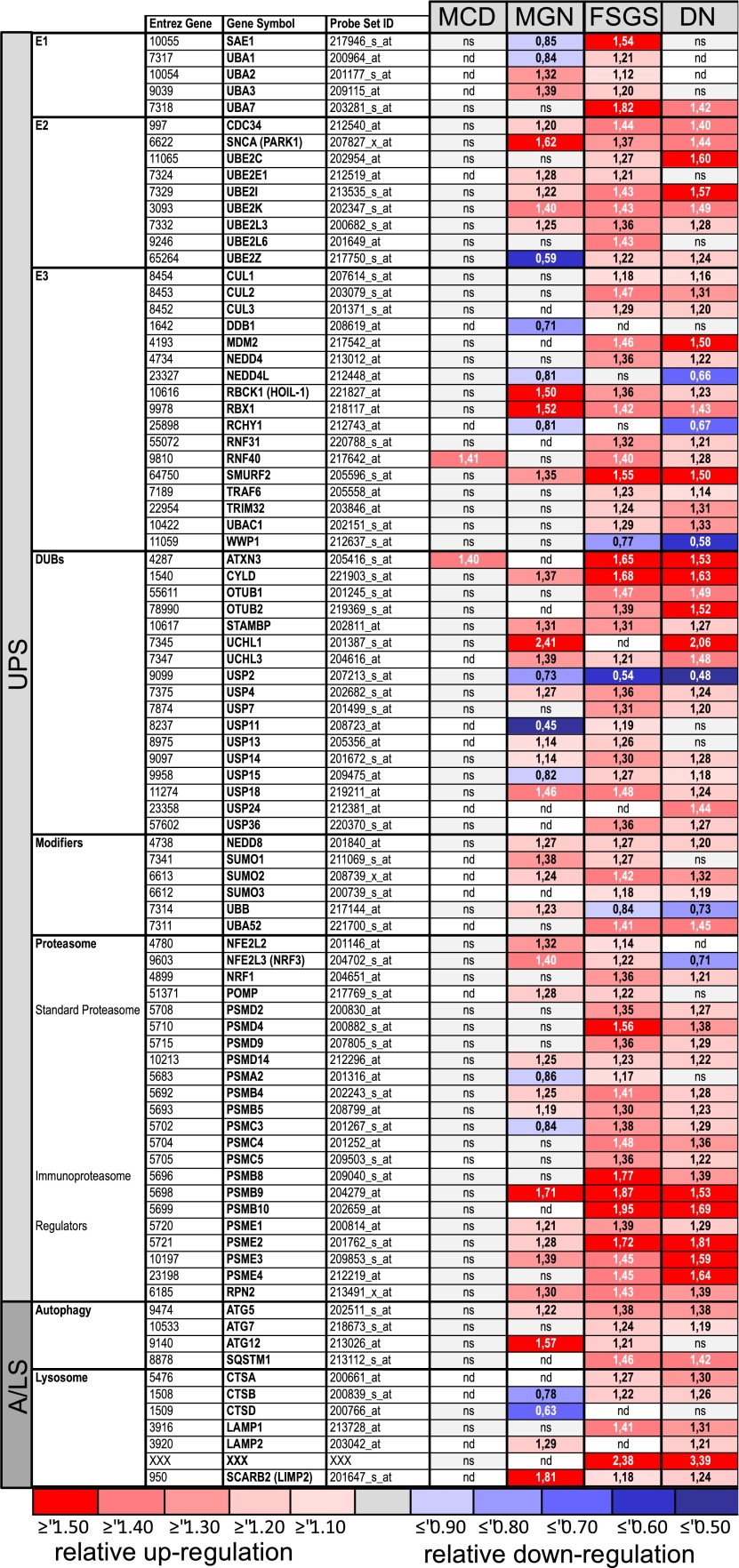
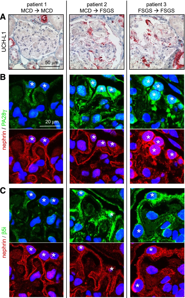
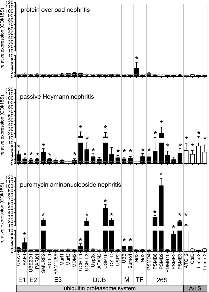
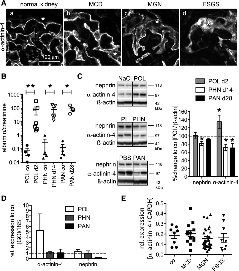
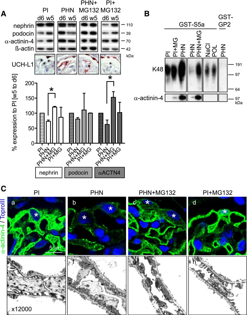
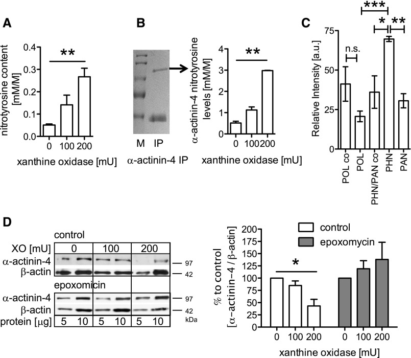
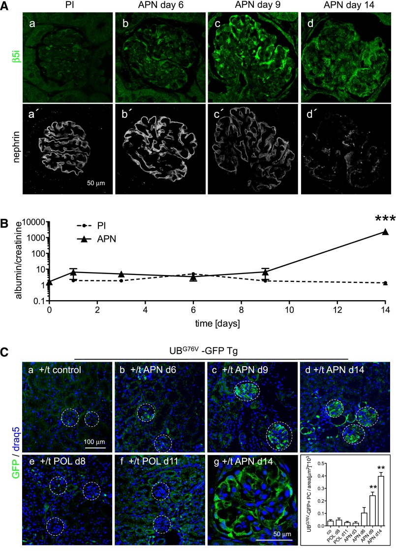
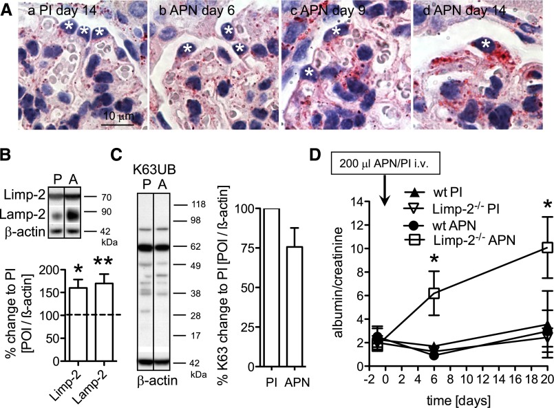
Podocytes are the key cells affected in nephrotic glomerular kidney diseases, and they respond uniformly to injury with cytoskeletal rearrangement. In nephrotic diseases, such as membranous nephropathy and FSGS, persistent injury often leads to irreversible structural damage, whereas in minimal change disease, structural alterations are mostly transient. The factors leading to persistent podocyte injury are currently unknown. Proteolysis is an irreversible process and could trigger persistent podocyte injury through degradation of podocyte-specific proteins. We, therefore, analyzed the expression and functional consequence of the two most prominent proteolytic systems, the ubiquitin proteasome system (UPS) and the autophagosomal/lysosomal system, in persistent and transient podocyte injuries. We show that differential upregulation of both proteolytic systems occurs in persistent human and rodent podocyte injury. The expression of specific UPS proteins in podocytes differentiated children with minimal change disease from children with FSGS and correlated with poor clinical outcome. Degradation of the podocyte-specific protein α-actinin-4 by the UPS depended on oxidative modification in membranous nephropathy. Notably, the UPS was overwhelmed in podocytes during experimental glomerular disease, resulting in abnormal protein accumulation and compensatory upregulation of the autophagosomal/lysosomal system. Accordingly, inhibition of both proteolytic systems enhanced proteinuria in persistent nephrotic disease. This study identifies altered proteolysis as a feature of persistent podocyte injury. In the future, specific UPS proteins may serve as new biomarkers or therapeutic targets in persistent nephrotic syndrome.


Figures

Figure 1.
Figure 1.
Proteolytic systems are upregulated in persistent human glomerular injury; 128 genes encoding UPS or A/LS components were selected on literature research, and their relative transcript expression levels were analyzed by microarrays in microdissected glomeruli from patients with biopsy-proven MCD (LD [n=18] versus MCD [n=5]), MGN (LD [n=14] versus MGN [n=21]), FSGS (LD [n=18] versus FSGS [n=10]), and DN (LD [n=18] versus DN [n=7]). Eighty-seven genes were above the cutoff. The cutoff was defined as (1) regulation in at least two diseases or (2) at least a 40% regulation, which means a fold change≥1.4 or ≤0.71. Heat maps of all genes with a significant regulation in MCD, MGN, FSGS, and/or DN are shown. Numbers represent fold changes compared with the respective controls (LDs). Note the difference in regulation between MCD and the more persistent glomerular injuries. DUBs, deubiquitinating enzymes; nd, not detected.
Figure 2.
Figure 2.
Expression of UPS proteins in podocytes during human nephrotic syndrome is a sign for the progression to ESRD. Renal biopsies of patients with the primary diagnosis of MCD (patients 1 and 2) or FSGS (patient 3) were stained for UPS proteins. (A) Deubiquitinating enzyme UCH-L1 (red). (B) Proteasome activator PA28γ (green; costained with nephrin [red]). (C) β5i unit of the immunoproteasome (green; costained with nephrin [red]). Patients 2 and 3 received the end diagnosis FSGS during clinical follow-up (→) and progressed to ESRD. Patient 1 went into remission and maintained the diagnosis of MCD. Note the increased expression of UPS proteins in patient 2 in podocytes with intact nephrin staining. *Podocyte nucleus.
Figure 3.
Figure 3.
Proteolytic systems are differentially upregulated in persistent rat glomerular injury. Relative glomerular expression of transcripts encoding for ubiquitin proteasome players (black bars: E1, ubiquitin activating enzymes; E2, ubiquitin binding enzymes; E3, ubiquitin ligating enzymes; DUB, deubiquitinating enzymes; M, modifiers [ubiquitin or ubiquitin-like proteins]; TF, transcription factors; 26S, proteasome subunits) or players involved in the autophagosomal/lysosomal degradation system in rats (A/LS; white bars). Transient podocyte injury was induced by BSA injection (protein overload; n=4; day 2). Persistent podocyte injury was induced by injection of FX1A serum (PHN; n=6; 18 days) or PAN (n=4; 4 weeks) as described in detail in Concise Methods. Values were normalized to 18S RNA as the housekeeping gene of the same preparations, and the relative expressions to controls were calculated. *P<0.05 was accepted as statistically significant to respective controls (dashed gray line=1). GOI, gene of interest.
Figure 4.
Figure 4.
Foot-process proteins, such as nephrin and α-actinin-4, are decreased in persistent human and rat glomerular injury. (A) Confocal micrographs of α-actinin-4 immunolocalization in renal biopsy specimens of control (a), MCD (b), MGN (c), or FSGS (d). *Podocyte cell body. Note disrupted α-actinin-4 staining in MGN and FSGS. (B–D) Transient podocyte damage was induced in rats through intraperitoneal BSA injection day 1, and persistent podocyte damage was induced through intravenous injection of FX1A serum day −1 and day 0 or intraperitoneal injection of puromycin amino nucleoside. (B) Urine albumin-to-creatinine ratio at the day of euthanization (day 2, POL control [NaCl] and POL; day 18, PHN control [PI] and PHN; week 4, PAN control [PBS] and PAN). *P<0.05; **P<0.01. (C, left) Western blotting for nephrin and α-actinin-4 from isolated glomeruli of POL rats on day 2, PHN rats on day 18, and PAN rats on day 28 after disease induction. (C, right) Densitometric analysis of nephrin and α-actinin-4 levels from two independent experiments with n=3–4 samples per condition. Values were normalized to β-actin of the same membrane and are expressed as percent change to control rats (co; dashed line). *P<0.05. POI, protein of interest. (D) Relative glomerular mRNA expression levels of nephrin and α-actinin-4 in POL glomeruli (n=6; day 2), PHN glomeruli (n=6; 14 days), or PAN glomeruli (n=6; 4 weeks) to respective controls. Values were normalized to 18S RNA as a housekeeping gene of the same preparations and are expressed as mean±SEM; no statistical significance was noted to respective controls (dashed gray line=1). (E) Relative expression levels of α-actinin-4 mRNA in microdissected human glomeruli of LD (co; n=8) or biopsy-proven MCD (n=14), MGN (n=29), and FSGS (n=9). Glyceraldehyde-3-phosphate dehydrogenase (GAPDH) mRNA was used as an internal housekeeper to adjust for unequal total mRNA content. GOI, gene of interest.
Figure 5.
Figure 5.
The UPS degrades foot-process proteins in persistent podocyte injury. (A) Inhibition of proteasomal degradation stabilizes nephrin, podocin, and α-actinin-4 in PHN rats. PHN was induced in rats day −1 and day 0 through intravenous injection of FX1A (PHN) or PI (control). On day 6, a unilateral nephrectomy was performed, and glomeruli were isolated. Proteasomal activity was inhibited with MG132 (60 μg/kg body wt) for 3 days before euthanization at week 5. (Top) Western blotting for nephrin, podocin, and α-actinin-4 from day 6- and week 5-isolated glomeruli from the same rat shows reduction of foot-process proteins in PHN rats, which is abolished by inhibition of the proteasome. β-Actin of the same membrane was used as a loading control. (Middle) Micrographs of UCH-L1 expression (red) as a surrogate marker of UPS upregulation in respective rats at week 5. *Podocytes. (Bottom) Densitometric analysis of percent changes of nephrin, podocin, and α-actinin-4 levels from week 5 to day 6 glomeruli of individual rats (n=2–3) compared with control rats (PI; striped bars). *P<0.05. (B) In a different set of experiments, K48-polyubiquitinated proteins were pulled down from glomerular lysates through the GST-S5a matrix. PHN was performed without uninephrectomy to provide sufficient glomerular protein per rat 28 days after disease induction with or without prior proteasomal inhibition for 2 weeks. Upper panel shows increased pull down of K48-polyubiquitinated proteins from 100 μg glomerular lysate in PHN rats and PHN rats with proteasomal inhibition by Western blotting for ubiquitin. After induction of transient podocyte injury in the model of protein overload day 2 (POL), no increased content of K48-polyubiquitinated proteins was noted to NaCl control-injected rats. GST-GP2 matrix was used to control for unspecific pull down (lane 8, long exposition). The same membrane was then subjected to Western blotting for K48-polyubiquitinated α-actinin-4 within the precipitate. Note the increased content of K48-polyubiquitinated α-actinin-4 in PHN rats (lane 3), which was further increased by proteasomal inhibition (lane 5). No K48-polyubiquitinated α-actinin-4 was detected in POL rats. Three independent experiments were performed. (C) Immunolocalization (a–d) of α-actinin-4 and electron micrographs (e–h) in PI and PHN rats 5 weeks after disease induction after a 3-day course of proteasomal inhibition. *Podocyte nucleus.
Figure 6.
Figure 6.
The UPS in human podocytes and rat persistent podocyte injury degrades damaged α-actinin-4. (A) Human podocytes were treated for 5 hours with different concentrations of XO and analyzed for total nitrotyrosine content as a marker for oxidative stress (n=3). (B) α-Actinin-4 protein was immunopurified from untreated and XO-treated podocytes and separated using SDS-PAGE followed by a coomassie staining (left; arrow, α-actinin-4). IP, immunoprecipitate; M, marker. (Right) The α-actinin-4 protein band was cut from the gel and analyzed for nitrotyrosine modifications using MS. A significant increase of nitrotyrosine modifications within the α-actinin-4 protein was seen in samples exposed to XO (n=2). (C) Immunopurified α-actinin-4 from glomerular lysates of PHN day 14 (n=2), PAN week 4 (n=3), and POL day 2 (n=2) treated rats and respective control rats (POL co, n=2; PHN/PAN co, n=3) was separated by SDS-PAGE and subjected to MS analysis for oxidative modifications. The amounts of the identified tryptic α-actinin-4 peptide M*TLGM*IW**TIILR (m/z=756.2) [M+2H]2+ with a dioxidized tryptophan (W**) and two oxidized methionines (M*) are shown relative to an unmodified α-actinin-4 peptide. (D, left) Western blotting for α-actinin-4 from protein lysates of human podocytes exposed to 5 hours of XO with or without pretreatment with the proteasome inhibitor epoxomicin (10 μM). Two concentrations of total protein lysates (5 and 10 μg) were loaded for reference. (D, right) Densitometric analysis of three to four independent experiments with n=1 sample per condition. Values were normalized to β-actin of the same membrane and are expressed as percent change to control (0 mU XO). *P<0.5; **P<0.01; ***P<0.001.
Figure 7.
Figure 7.
Proteasomal proteolytic capacity is overwhelmed before the development of proteinuria in persistent podocyte injury. (A) Confocal images of staining for the immunoproteasome subunit β5i (a–d; green) and nephrin (a′–d′; white) days 6, 9, and 14 after induction of APN or in a PI-injected control mouse day 14 show an early expression of β5i in podocytes of APN mice before disrupted nephrin staining. (B) Time course of development of albuminuria measured by ELISA. Values were normalized against urine creatinine of respective urine samples. ***P<0.001. (C) Confocal images of staining for GFP (green) in UBG76V-GFP transgenic mice 14 days after injection of PI or APN serum show early accumulation of UBG76V-GFP in podocytes of APN mice. Induction of reversible podocyte injury in the model of POL did not result in UBG76V-GFP accumulation in podocytes during (day 8; e) and after (day 11; f) intraperitoneal administration of BSA for 7 subsequent days. (g) Glomerular close-up view of an UBG76V-GFP transgenic mouse day 14 after APN induction. (h) GFP-positive podocytes were counted in 20 glomeruli and normalized against glomerular area in the different mouse groups. **P<0.001
Figure 8.
Figure 8.
The autophagosomal/lysosomal system compensates for impaired UPS function. APN was induced day 0 by intravenous injection of 300 μl sheep APN or unspecific sheep PI (control). (A) Staining for the lysosomal protein Limp-2 in APN mice and a PI-injected control mouse shows a significant accumulation of lysosomes in podocytes on day 14 in APN. (B) Lysosomal Limp-2 and lysososmal-associated membrane protein-2 (LAMP-2) levels are increased in isolated glomeruli of APN mice (A) relative to PI (P=1.00; dotted line) day 14 mice treated by Western blotting and densitometric analysis. Values were normalized to β-actin of the same membrane and are expressed as percent change to PI mice (n=3–6 animals per time point and condition). (C) K63-polyubiquitinated proteins from glomerular lysates of PI and APN mice were quantified day 14 by Western blotting and densitometric analysis (n=3 mice per condition). (D) Time course of urine albumin-to-creatinine ratio in Limp-2–deficient mice and control littermates after induction of mild APN through intravenous (i.v.) injection of 200 μl APN or PI serum (n=3–4 for all conditions and time points). *P<0.05; **P<0.01.

References

    1. Guan N, Ding J, Zhang J, Yang J: Expression of nephrin, podocin, alpha-actinin, and WT1 in children with nephrotic syndrome. Pediatr Nephrol 18: 1122–1127, 2003 - PubMed
    1. Huh W, Kim DJ, Kim MK, Kim YG, Oh HY, Ruotsalainen V, Tryggvason K: Expression of nephrin in acquired human glomerular disease. Nephrol Dial Transplant 17: 478–484, 2002 - PubMed
    1. Luimula P, Sandström N, Novikov D, Holthöfer H: Podocyte-associated molecules in puromycin aminonucleoside nephrosis of the rat. Lab Invest 82: 713–718, 2002 - PubMed
    1. Ciechanover A, Orian A, Schwartz AL: The ubiquitin-mediated proteolytic pathway: Mode of action and clinical implications. J Cell Biochem Suppl 34: 40–51, 2000 - PubMed
    1. Hochstrasser M: Biochemistry. All in the ubiquitin family. Science 289: 563–564, 2000 - PubMed

Publication types

MeSH terms