. 2014 Aug;25(8):1669-78.
doi: 10.1681/ASN.2013101125. Epub 2014 Apr 10.

Human cytomegalovirus inhibits erythropoietin production

Affiliations

Human cytomegalovirus inhibits erythropoietin production

Lynn M Butler et al. J Am Soc Nephrol. 2014 Aug.

Abstract

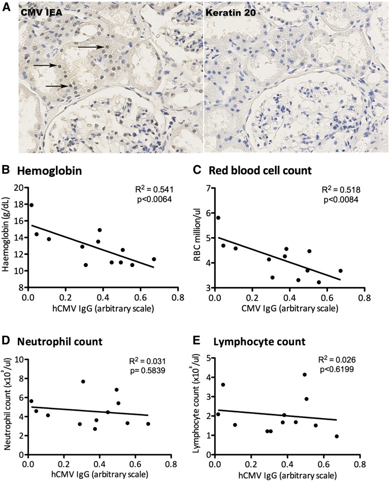
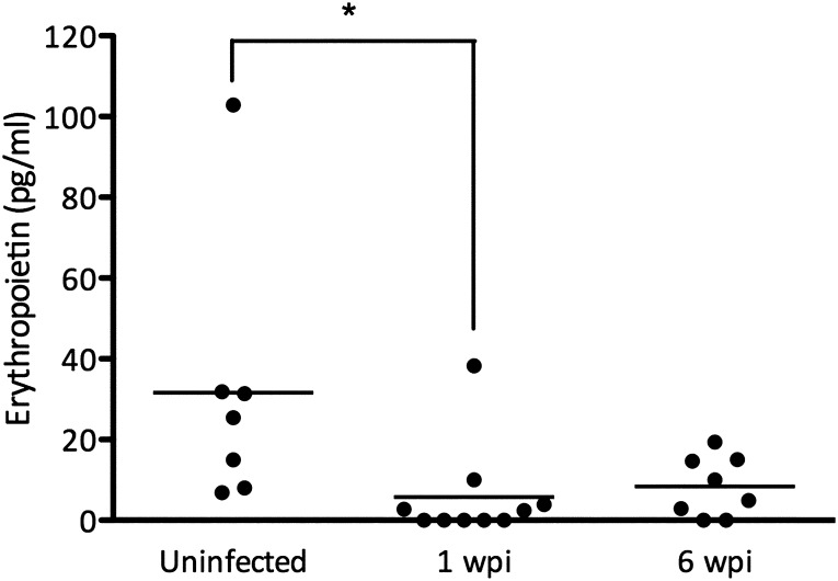
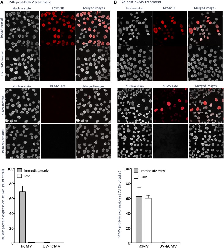
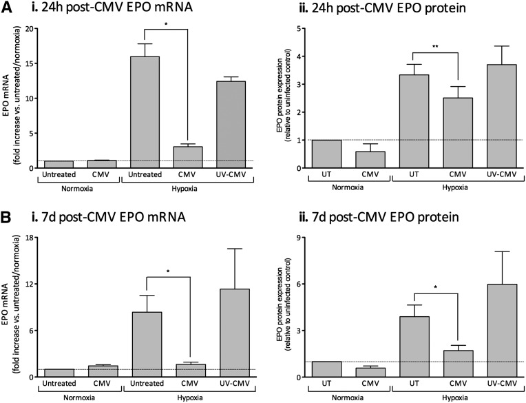
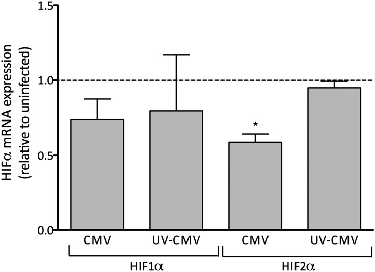
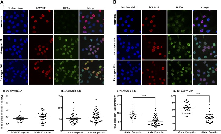
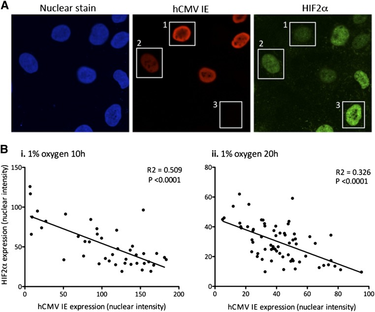
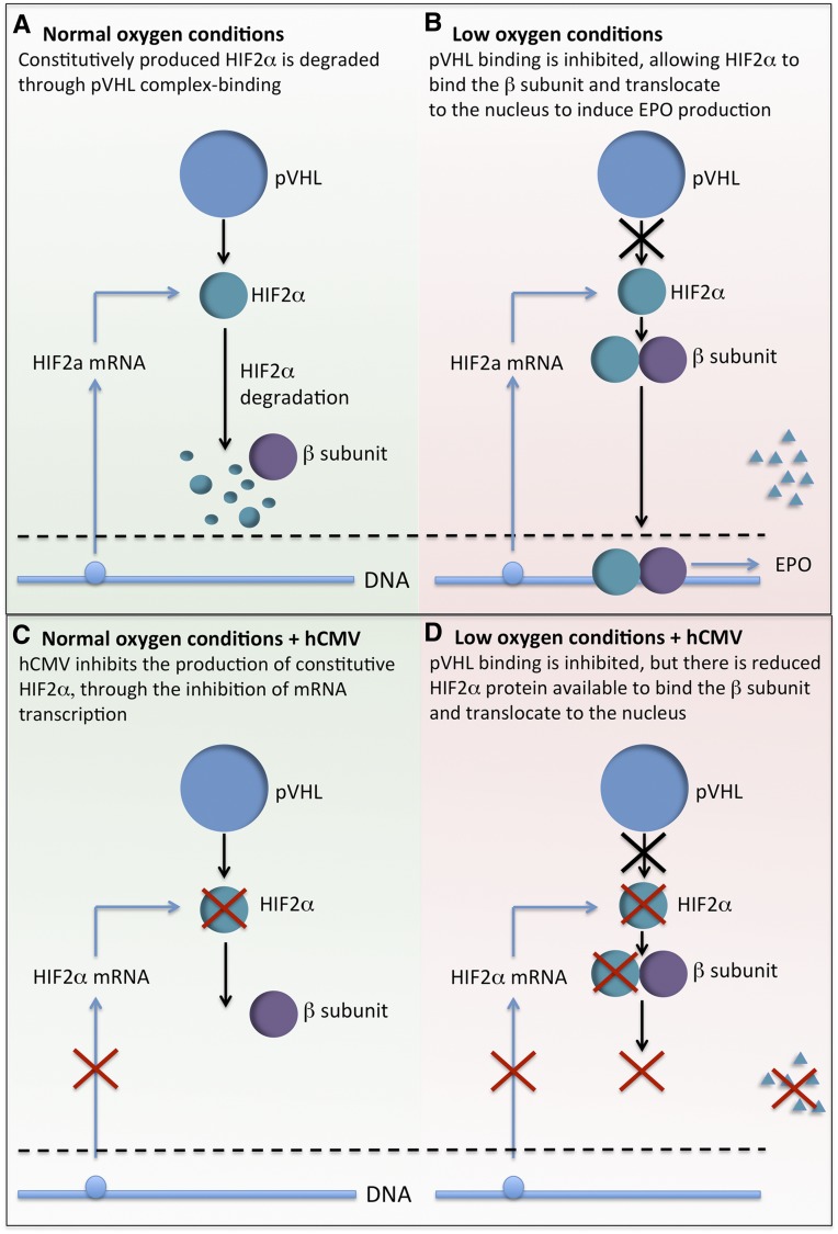
Anemia is a feature of CKD and a complication of renal transplantation, often caused by impaired production of erythropoietin. The kidney is a target organ for human cytomegalovirus (hCMV) in such patients, but it is not known whether hCMV effects erythropoietin production. We found that kidneys from patients with CKD were positive for hCMV protein and that blood levels of hCMV IgG inversely correlated with red blood cell count. In mice, systemic murine cytomegalovirus infection decreased serum erythropoietin levels. In human erythropoietin-producing cells, hCMV inhibited hypoxia-induced expression of erythropoietin mRNA and protein. hCMV early gene expression was responsible, as ultraviolet-inactivated virus had no effect and valganciclovir treatment showed that late gene expression was nonessential. Hypoxia-induced gene transcription is controlled by the transcription factors hypoxia-inducible transcription factor (HIF)-1α and HIF2α, which are constitutively produced but stable only under low oxygen conditions. We found that hCMV inhibited constitutive production of HIF2α mRNA. HIF2α is thought to be the master regulator of erythropoietin transcription. Single-cell analysis revealed that nuclear accumulation of HIF2α was inhibited in hCMV-infected cells, and the extent of inhibition correlated with hCMV protein expression. Our findings suggest that renal hCMV infection could induce or exacerbate anemia in patients.


Figures

Figure 1.
Figure 1.
Kidney tissue biopsy specimens from patients with CKD are positive for hCMV, and blood hCMV IgG levels inversely correlate with red blood cell count. (A) Biopsy specimens from CKD were stained for hCMV IE proteins; arrows highlight examples of positive nuclear staining. Images were captured using a magnification factor of ×40. (B–E) Correlation between hCMV IgG levels and serum hemoglobin (B), red blood cell count (C), neutrophil count (D), and lymphocyte count (E).
Figure 2.
Figure 2.
Murine CMV reduces baseline EPO levels in mice. BALB/c mice were left untreated or systemically infected with mCMV by intraperitoneal injection. Circulating EPO levels were determined 1 or 6 weeks after infection (*P<0.05, one-way ANOVA).
Figure 3.
Figure 3.
HEPCs are susceptible to hCMV infection, and ultraviolet (UV) inactivation efficiently prevents viral protein expression. Cells were treated with hCMV or ultraviolet-inactivated hCMV at an MOI of 5. Expression of hCMV IE or late antigen was determined by immunocytochemistry and confocal microscopy at 24 hours (A) or 7 days (B) after treatment. Images are representative of three independent experiments. Images were captured using a magnification factor of ×40. Data are mean±SEM from three to eight independent experiments.
Figure 4.
Figure 4.
hCMV inhibits HEPC EPO production under conditions of hypoxia. Cells were treated with hCMV or ultraviolet (UV)-inactivated hCMV at an MOI of 5 and cultured for 24 hours (A) or 7 days (B) before exposure to 1% O2 or maintenance at normoxia for 18–20 hours before analysis of EPO mRNA production (i) or EPO supernatant protein level (ii) (both plotted relative to the untreated control under normoxic conditions). Data are mean±SEM from three to seven independent experiments. *P<0.05; **P< 0.01, versus untreated (UT).
Figure 5.
Figure 5.
hCMV inhibits constitutive HIF2α mRNA expression in HEPCs. Cells were treated with hCMV or ultraviolet (UV)-inactivated hCMV at an MOI of 5 and cultured for 24 hours before exposure to 1% O2 for 18–20 hours. Levels of HIF1α and HIF2α mRNA were determined by qPCR. Data are mean±SEM from three to four independent experiments. *P<0.05, versus untreated.
Figure 6.
Figure 6.
hCMV inhibits hypoxia-induced nuclear translocation of HIF2α. HEPCs were treated with hCMV at a MOI of 1 and cultured for 24 hours before exposure to 1% O2, or maintenance at normoxia, for 10 or 20 hours before fixation and staining for HIF1α or HIF2α, and hCMV IE protein expression. Levels of HIF1α or HIF2α, and hCMV IE were observed by confocal microscopy (Ai and Bi). Images were captured using a magnification factor of ×40. Images were analyzed for nuclear fluorescence in the hCMV-negative and -positive populations following exposure to 1% O2 for 10 hours (Aii and Bii) or 20 hours (Aiii and Biii). ***P<0.001.
Figure 7.
Figure 7.
Expression of hCMV IE negatively correlates with levels of nuclear HIF2α. HEPCs were treated with hCMV at a MOI of 1 and cultured for 24 hours before exposure to 1% O2, or maintenance at normoxia, for either 10 or 20 hours before observation of HIF2α IE protein confocal microscopy. (A) Boxes highlight cells that express (1) high IE protein, low HIF2α; (2) moderate IE protein, moderate HIF2α; and (3) no IE protein, high HIF2α. Images are representative of three independent experiments. Images were captured using a magnification factor of ×63. (B) hCMV-positive cells were analyzed for nuclear intensity of IE protein and HIF2α following exposure to 1% O2 for 10 hours (i) or 20 hours (ii). Data are from three independent experiments.
Figure 8.
Figure 8.
Working model of the effect of hCMV on EPO production. (A) Under conditions of normal oxygen, HIF2α is constitutively produced, bound to proteasomal degradation by the von Hippel-Lindau tumor suppressor (pVHL), and targeted for degradation. (B) Under conditions of low oxygen, pVHL does not bind to HIF2α, so it is spared from degradation. It can then bind to the β-subunit, translocate to the nucleus, and induce the transcription EPO. (C) Under conditions of normal oxygen, hCMV inhibits the constitutive production of HIF2α, but there is no noticeable effect because the protein is usually degraded. (D) Under conditions of low oxygen, the hCMV-induced inhibition of HIF2α protein production results in being less spared from degradation, reduced binding to the β-subunit, and thus reduced transcription of EPO.

Comment in

References

    1. Lorenz M, Kletzmayr J, Perschl A, Furrer A, Hörl WH, Sunder-Plassmann G: Anemia and iron deficiencies among long-term renal transplant recipients. J Am Soc Nephrol 13: 794–797, 2002 - PubMed
    1. Shah N, Al-Khoury S, Afzali B, Covic A, Roche A, Marsh J, Macdougall IC, Goldsmith DJ: Posttransplantation anemia in adult renal allograft recipients: Prevalence and predictors. Transplantation 81: 1112–1118, 2006 - PubMed
    1. Winkelmayer WC, Chandraker A, Alan Brookhart M, Kramar R, Sunder-Plassmann G: A prospective study of anaemia and long-term outcomes in kidney transplant recipients. Nephrol Dial Transplant 21: 3559–3566, 2006 - PubMed
    1. Vanrenterghem Y, Ponticelli C, Morales JM, Abramowicz D, Baboolal K, Eklund B, Kliem V, Legendre C, Morais Sarmento AL, Vincenti F: Prevalence and management of anemia in renal transplant recipients: A European survey. Am J Transplant 3: 835–845, 2003 - PubMed
    1. Vanrenterghem Y: Anaemia after renal transplantation. Nephrol Dial Transplant 19[Suppl 5]: V54–V58, 2004 - PubMed

Publication types

MeSH terms