Review
. 2015 Apr;21(2):169-84.
doi: 10.1177/1073858414530512. Epub 2014 Apr 10.

Microglial dynamics and role in the healthy and diseased brain: a paradigm of functional plasticity

Affiliations
Review

Microglial dynamics and role in the healthy and diseased brain: a paradigm of functional plasticity

Diego Gomez-Nicola et al. Neuroscientist. 2015 Apr.

Abstract

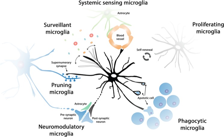
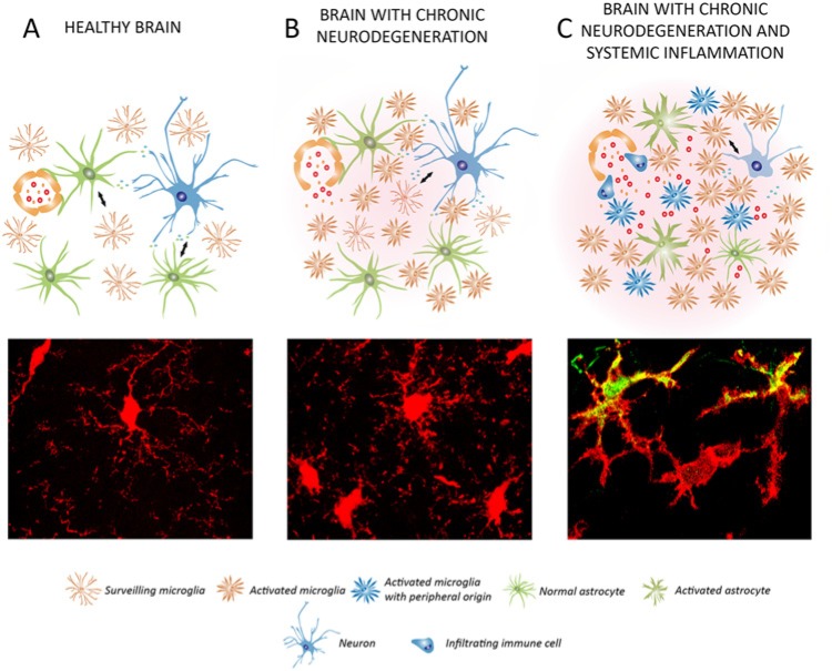
The study of the dynamics and functions of microglia in the healthy and diseased brain is a matter of intense scientific activity. The application of new techniques and new experimental approaches has allowed the identification of novel microglial functions and the redefinition of classic ones. In this review, we propose the study of microglial functions, rather than their molecular profiles, to better understand and define the roles of these cells in the brain. We review current knowledge on the role of surveillant microglia, proliferating microglia, pruning/neuromodulatory microglia, phagocytic microglia, and inflammatory microglia and the molecular profiles that are associated with these functions. In the remodeling scenario of microglial biology, the analysis of microglial functional states will inform about the roles in health and disease and will guide us to a more precise understanding of the multifaceted roles of this never-resting cells.

Keywords: microglia; neuroinflammation; neuromodulation; phagocytosis; proliferation; surveillance; synaptic pruning.


Conflict of interest statement

Declaration of Conflicting Interests: The author(s) declared no potential conflicts of interest with respect to the research, authorship, and/or publication of this article.

Figures

Figure 1.
Figure 1.
Functional states of microglia in the healthy brain. The population of microglial cells is maintained by self-renewal, without the contribution of bone-marrow-derived progenitors. Surveillant microglial cells constantly scan the brain microenvironment, in order to detect minor perturbations of CNS homeostasis. Surveillant microglia can, for example, detect the presence of neurotoxic substances or inflammatory mediators from the systemic circulation, being in close communication with the blood-brain barrier (systemic sensing microglia). Phagocytic microglia can detect and quickly remove damaged or dying neurons, preventing the damage to neighboring cells and helping maintain the high turnover of specific cell populations (i.e., neural precursor cells). The phagocytic capacity of microglia is particularly important in development (pruning microglia), when they can contribute to the removal of supernumerary synapses in certain neuronal pathways. Moreover, it has been suggested that microglia can have a direct or indirect modulatory role at the synapse, influencing neuronal activity (neuromodulatory microglia).
Figure 2.
Figure 2.
Dynamics and functions of microglia in chronic neurodegenerative diseases. (A) In the normal brain, the microglial population has a surveillant phenotype, maintaining homeostasis. The microglia population is maintained by self-renewal, while the perivascular macrophages can be renewed by bone-marrow-derived progenitors. In Alzheimer’s disease (AD) microglia proliferate and accumulate around plaques of amyloid β (Aβ), participating in the attempted removal of the misfolded protein. Perivascular macrophages have a more efficient phagocytic activity than microglial cells in AD. In AD, the microglial population is increased without a contribution from bone-marrow-derived cells. Microglia are expanded and activated during the course of amyotrophic lateral sclerosis (ALS), without a contribution from circulating progenitors. In prion disease, the microglial population is expanded dramatically by local proliferation (B; BrdU+), being primed to give an exaggerated response to systemic inflammatory events. Little evidence is available about the expansion/renewal of the microglial population during Parkinson’s or Huntington’s disease, or the dominant inflammatory phenotype. In general, the microglial population does not generate a uniform response and a diverse inflammatory prolife can coexist during disease (C; CD11c+ vs. CD11c microglia). For all the neurodegenerative diseases considered, little evidence is available about the possible contribution of perivascular macrophages (D; CD163+ CCR2+) to the expansion/renewal of the microglial population (D; CD163 CCR2), although both populations have different activation and proliferation patterns. (B-D) Representative examples evidenced in prion disease, detecting microglial cells by the transgenic expression of EGFP under the c-fms promoter.
Figure 3.
Figure 3.
Impact of systemic inflammation on the progression of chronic neurodegeneration: microglial priming. Schematic representation of the cross-talk of microglial cells with neurons and astrocytes in the healthy brain (A), during chronic neurodegeneration (B), and when chronic neurodegeneration is combined with a systemic inflammatory event (C). (A) In the healthy brain, surveillant microglia maintain the brain homeostasis and are renewed by local proliferation. Astrocytes and microglia communicate with neurons to support their function and survival, among other functions (see Fig. 1). (B) In chronic neurodegeneration, microglia activate an inflammatory program and become primed. The microglial population is expanded mainly by local proliferation. Astrocytes lose control of the blood-brain barrier and inflammatory mediators and cells enter into the brain. Neurons undergo a progressive but limited damage. (C) When a systemic inflammatory event is combined with chronic neurodegeneration, primed microglia are further activated and damage endangered neurons, accelerating the pathology. The microglial population can be supplemented by bone-marrow-derived cells. Astrocytes become activated and further contribute to neuronal damage. (A-C) Microglial cells (red) exemplifying the different conditions are shown at the bottom. In C, primed microglial cells are shifted to a pro-inflammatory phenotype, expressing IL1β (green). The legend for the different cell types and phenotypes is provided at the bottom.

References

    1. Ajami B, Bennett JL, Krieger C, McNagny KM, Rossi FM. 2011. Infiltrating monocytes trigger EAE progression, but do not contribute to the resident microglia pool. Nat Neurosci 14(9):1142–9. - PubMed
    1. Ajami B, Bennett JL, Krieger C, Tetzlaff W, Rossi FM. 2007. Local self-renewal can sustain CNS microglia maintenance and function throughout adult life. Nat Neurosci 10(12):1538–43. - PubMed
    1. Akiyama H, Barger S, Barnum S, Bradt B, Bauer J, Cole GM, and others. 2000. Inflammation and Alzheimer’s disease. Neurobiol Aging 21(3):383–421. - PMC - PubMed
    1. Akiyama H, Nishimura T, Kondo H, Ikeda K, Hayashi Y, McGeer PL. 1994. Expression of the receptor for macrophage colony stimulating factor by brain microglia and its upregulation in brains of patients with Alzheimer’s disease and amyotrophic lateral sclerosis. Brain Res 639(1):171–4. - PubMed
    1. Alliot F, Godin I, Pessac B. 1999. Microglia derive from progenitors, originating from the yolk sac, and which proliferate in the brain. Brain Res Dev Brain Res 117(2):145–52. - PubMed

Publication types