. 2014 Apr 10;10(4):e1004027.
doi: 10.1371/journal.ppat.1004027. eCollection 2014 Apr.

Complement-related proteins control the flavivirus infection of Aedes aegypti by inducing antimicrobial peptides

Affiliations

Complement-related proteins control the flavivirus infection of Aedes aegypti by inducing antimicrobial peptides

Xiaoping Xiao et al. PLoS Pathog. .

Abstract

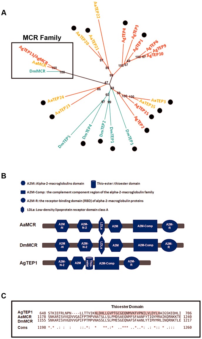
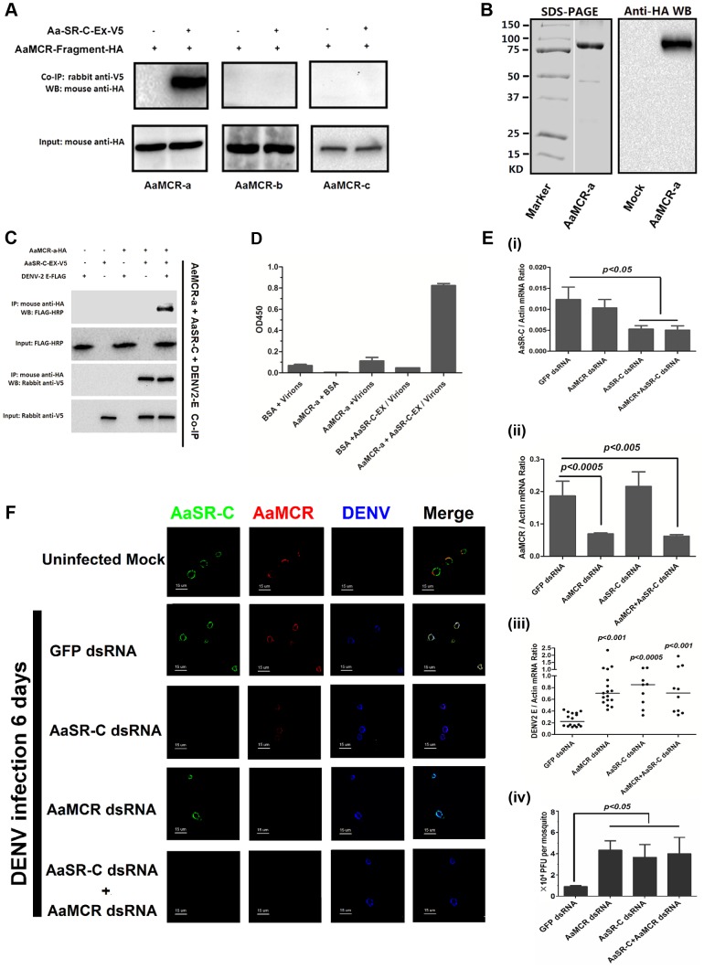
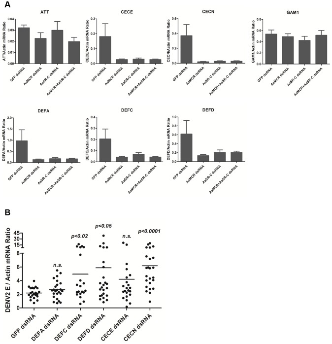
The complement system functions during the early phase of infection and directly mediates pathogen elimination. The recent identification of complement-like factors in arthropods indicates that this system shares common ancestry in vertebrates and invertebrates as an immune defense mechanism. Thioester (TE)-containing proteins (TEPs), which show high similarity to mammalian complement C3, are thought to play a key role in innate immunity in arthropods. Herein, we report that a viral recognition cascade composed of two complement-related proteins limits the flaviviral infection of Aedes aegypti. An A. aegypti macroglobulin complement-related factor (AaMCR), belonging to the insect TEP family, is a crucial effector in opposing the flaviviral infection of A. aegypti. However, AaMCR does not directly interact with DENV, and its antiviral effect requires an A. aegypti homologue of scavenger receptor-C (AaSR-C), which interacts with DENV and AaMCR simultaneously in vitro and in vivo. Furthermore, recognition of DENV by the AaSR-C/AaMCR axis regulates the expression of antimicrobial peptides (AMPs), which exerts potent anti-DENV activity. Our results both demonstrate the existence of a viral recognition pathway that controls the flaviviral infection by inducing AMPs and offer insights into a previously unappreciated antiviral function of the complement-like system in arthropods.


Conflict of interest statement

The authors have declared that no competing interests exist.

Figures

Figure 1
Figure 1. Comparison of the functional domains and phylogenetic analysis of insect thioester-containing proteins (iTEPs).
(A) Unrooted phylogenetic tree of iTEPs. The tree was constructed using the neighbour-joining (NJ) method based on the alignment of 23 iTEP protein sequences. The bootstrap values of 500 replicates (%) are indicated on the branch nodes. Anopheles gambiea (Ag), Aedes Aegypti (Aa) and Drosophila melanogaster (Dm) are indicated with red, yellow and green, respectively. Period represents the iTEPs with thioester domain. (B) Schematic representation of AaMCR, DmMCR and AgTEP1. The functional modules of MCRs and TEPs were predicted using the SMART (http://smart.embl-heidelberg.de/smart/set_mode.cgi?GENOMIC=1) and Pfam websites (http://pfam.sanger.ac.uk/). (C) Alignment of the sequence of thioester domain using CLUSTAL-X. The shadowed sequence is the predicted thoiester domain. Asterisk indicates the identical residues in all sequences of the alignment; colon indicates the conserved substitutions; period indicates the semi-conserved substitutions.
Figure 2
Figure 2. AaMCR restricts flaviviral infections.
(A-B) Knockdown of the AaMCR gene. The mosquitoes were microinjected with AaMCR or GFP dsRNA. The inoculated mosquitoes were sacrificed, and AaMCR abundance was assessed via qPCR (A) and immuno-blotting with an AaMCR antibody (B) at 6 days post microinjection. 50 μg of protein from mosquito lysates was loaded into each lane (B). (C-G) Silencing of AaMCR enhanced DENVs and YFV (17D) infections in A. aegypti. 10 MID50 DENVs or YFV was inoculated at 3 days post-AaMCR silencing. The viral load was assessed at 6 days post-infection through qPCR and normalized against A. aegypti actin (AAEL011197). The primers and probes used for qPCR are described in Table S2. The experiment was repeated three times with similar results. One dot represents 1 mosquito and the horizontal line represents the median of the results. The data were analyzed statistically by the non-parametric Mann-Whitney test. (H) Immuno-blockade of AaMCR enhanced the DENV-2 infection of A. aegypti. The AaMCR-N antibody, in 10-fold serial dilutions, was premixed with 10 MID50 DENV-2 for thoracic co-microinjection. The treated mosquitoes were sacrificed to examine the viral load at 3 (i) and 6 (ii) days post-infection via qPCR and normalized against A. aegypti actin. The results were pooled from 3 independent experiments. One dot represents 1 mosquito and the horizontal line represents the median of the results. The data were analyzed statistically using the non-parametric Mann-Whitney test. (I) The expression of AaMCR fragment peptides in Drosophila S2 cells. Three fragments of the AaMCR (a, 30–601 aa; b, 590–1,240 aa; c, 1,200–1,793 aa) with a C-terminal HA tag were cloned into the pMT/BiP/V5-His A vector and expressed in the S2 cell supernatant. The supernatant from empty vector-transfected S2 cells was used as a mock. The recombinant peptides were detected with an anti-HA antibody via western blotting. (J) The fragments of AaMCR do not directly interact with DENV-2 E protein in ELISA. The cell supernatant, including the AaMCR fragments were collected at 48 hrs after transfection. The DENV-2 E protein was expressed and purified from a Drosophila expression system. Each plate well was coated with 2 ug of DENV-2 E. The interaction was determined using an anti-HA antibody. The empty DNA vector-transfected S2 cell supernatant was used as a negative control. The data are expressed as the mean ± standard error from 3 independent experiments.
Figure 3
Figure 3. The role of CCP domain-containing proteins in flaviviral infection.
Each CCP gene was silenced through thoracic microinjection of dsRNA and the effect on the viral burden was assessed on 6 days post-DENV-2 (A) or YFV (B) infection. GFP dsRNA-inoculated mosquitoes served as mock controls. The viral loads were measured via qPCR, and normalized against A. aegypti actin. One dot represents 1 mosquito and the horizontal line represents the median. The data were analyzed statistically using the non-parametric Mann-Whitney test. The results were combined from 2 independent experiments.
Figure 4
Figure 4. A Scavenger receptor-C with CCP domains recognizes DENV-2 in vitro and in vivo.
(A) Percentage of amino acid identity of insect SR-Cs. (B) Schematic representation of SR-Cs in A. aegypti and D. melanogaster. The functional modules were predicted in SMART (http://smart.embl-heidelberg.de/smart/set_mode.cgi?GENOMIC=1) and Pfam websites (http://pfam.sanger.ac.uk/). (C) Expression and purification of AaSR-C from Drosophila S2 cells. The extracellular region (AaSR-C-Ex) or full length (AaSR-C-Full) AaSR-C was cloned into the pMT/BiP/V5-His-A expression vector. The recombinant plasmids were transfected into Drosophila S2 cells, and their expression was probed using an anti-V5 mAb. The supernatant or lysates from mock-transfected S2 cells was used as the mock control (Left panel). Recombinant AaSR-C-Ex, produced in Drosophila cells, was purified using an Ni-His column (Right panel). (D) AaSR-C-Ex interacted with DENV-2 E proteins in co-immunoprecipitation assay. Purified AaSR-C-Ex (V5) and DENV-2 E (FLAG) were used to investigate the interaction of the proteins. Control rabbit IgG was used as a mock control to exclude non-specific interactions. The protein complex was pulled down with a rabbit anti-FLAG antibody and detected using a mouse anti-V5 antibody. We reproduced the experiment 3 times. (E) AaSR-C-Ex captured DENV-2 virions in an ELISA. Binding was probed using the flavivirus E mAb 4G2. The data are presented as the mean ± standard error. The experiment was reproduced 3 times. (F) AaSR-C bound DENV-2 virions on the cell surface. A Cu2+-inducible stable S2 cell line was generated to express the full-length AaSR-C. DENV-2 virions were incubated with AaSR-C expressing cells at 4°C for 1 hr. Non-induced stable cells and empty vector-transfected S2 cells containing virions served as the mock control groups. The interaction between AaSR-C and DENV was measured through flow cytometry. The DENV virions were stained using the flaviviral E mAb 4G2 and anti-mouse IgG Alexa-488; AaSR-C was probed using a Myc mAb and anti-rabbit IgG Phycoerythrin (PE). The data was analyzed using FlowJo software. The presented data was representative of 3 independent experiments with similar results. (G) The in vivo association between AaSR-C and DENV-2 in A.aegypti hemocytes. Hemolymph was collected from uninfected mosquitoes, AaSR-C silenced infected mosquitoes and GFP dsRNA treated infected mosquitoes to undergo immunofluorescence staining. AaSR-C was stained with anti-rabbit IgG Alexa-488 (Green), and the DENV-2 E protein was identified using anti-mouse IgG Alexa-546 (Red). Nuclei were stained blue with To-Pro-3 iodide (Blue). Images were examined using a Zeiss LSM 780 meta confocal 63×objective lens.
Figure 5
Figure 5. AaMCR and AaSR-C function in a pathway that opposes DENV-2 infection.
(A) The interaction between 3 AaMCR fragments and AaSR-C in co-IP assays. Three AaMCR gene fragments were cloned into the pMT/BiP/V5-His A vector. The recombinant plasmids were transiently transfected into S2 cells. The cell supernatant was used for investigation of the AaMCR/AaSR-C interaction. The protein complex was pulled down with a rabbit anti-V5 antibody and probed using a mouse anti-HA antibody. The experiment was reproduced 3 times. (B) Expression and purification of AaMCR-a in Drosophila S2 cells. The purified AaMCR-a was separated through SDS-PAGE (Left Panel) and detected with an anti-HA antibody via western blotting (Right Panel). The supernatant from empty vector-transfected S2 cells was used as the mock control. (C) AaSR-C-Ex acted as an adaptor in the interaction between the AaMCR-a and DENV-2 E proteins. The purified AaSR-C-Ex, AaMCR-a and DENV-2 E proteins were mixed and pulled down with a mouse anti-HA antibody (AaMCR-a) and detected using a rabbit anti-V5 antibody (AaSR-C) and anti-FLAG-HRP antibody (DENV-2 E). The experiment was repeated 3 times with similar results. (D) AaSR-C-Ex connected AaMCR-a to DENV-2 virions. Purified AaMCR-a or BSA was pre-coated into the ELISA plate wells. DENV-2 virions either mixed with AaSR-C-Ex or without AaSR-C-Ex were added to the protein-coated wells. The signal was detected using the flavivirus E mAb 4G2. The data are expressed as the mean ± standard error. The experiment was reproduced by 3 times with similar results. (E) Double knockdown of AaMCR and AaSR-C showed similar effects on DENV-2 infection to individual knockdown. Both AaSR-C (i) and AaMCR (ii) were knocked down using a dsRNA mixture in the AaSR-C/AaMCR co-silenced group. DENV-2 replication and the numbers of infectious DENV-2 virions in the mosquitoes were measured via qPCR (iii) and plaque assays (iv). Statistical analysis was performed using the non-parametric Mann-Whitney test. The data on gene silencing (i, ii) and from plaque assays (iv) are expressed as the mean ± standard error. The horizontal line depicts the median (iii). Each dot represents an individual mosquito. The result was representative of 3 independent experiments. (F) Immunostaining of AaMCR, AaSR-C and DENV-2 in A. aegypti hemocytes. The hemocytes were dissected from uninfected mosquitoes, AaMCR and/or AaSR-C silenced infected mosquitoes and GFP dsRNA treated infected mosquitoes at 6 days post-infection. AaSR-C was stained with anti-rabbit IgG Alexa-488 (Green); AaMCR was probed using anti-mouse IgG Alexa-546 (Red); the DENV-2 E protein was probed with DENV-2 human antiserum (purified IgG) and anti-human IgG Alexa-633 (Blue). Images were examined using a Zeiss LSM 780 meta confocal 63×objective lens.
Figure 6
Figure 6. The AaMCR/AaSR-C pathway induces antimicrobial peptides production to control dengue infection.
(A) Effects of AaMCR and AaSC-R knockdown on the expression of dengue-induced antimicrobial peptides. AaSR-C and AaMCR were knocked down individually or simultaneously through thoracic microinjection of dsRNA. Three days later, the gene-silenced mosquitoes were infected with (1,000 MID50) of DENV-2, and the AMP mRNA expression was assessed at 6 h post-infection via qPCR. The qPCR primers are provided in Table S2. The data is expressed as the mean± standard error of the results. The experiment was repeated 3 times. (B) Silencing of AMPs enhanced DENV-2 infection in A. aegypti. The 5 AaMCR/AaSR-C-regulated AMPs were knocked down through thoracic microinjection of dsRNA. DENV-2 (10 MID50) was inoculated at 3 days post AMP silencing. The viral load was assessed at 6 days post-infection via qPCR and normalized against A. aegypti actin (AAEL011197). The primers and probes used for qPCR are described in Table S2. The result was pooled from 3 independent experiments. One dot represents 1 mosquito and the horizontal line represents the mean value of the results. The data was analyzed statistically using the non-parametric Mann-Whitney test.

References

    1. Weaver SC, Barrett AD (2004) Transmission cycles, host range, evolution and emergence of arboviral disease. Nat Rev Microbiol 2: 789–801. - PMC - PubMed
    1. Ganesan S, Aggarwal K, Paquette N, Silverman N (2011) NF-kappaB/Rel proteins and the humoral immune responses of Drosophila melanogaster . Curr Top Microbiol Immunol 349: 25–60. - PMC - PubMed
    1. Shishido SN, Varahan S, Yuan K, Li X, Fleming SD (2012) Humoral innate immune response and disease. Clin Immunol 144: 142–158. - PMC - PubMed
    1. Levashina EA, Moita LF, Blandin S, Vriend G, Lagueux M, et al. (2001) Conserved role of a complement-like protein in phagocytosis revealed by dsRNA knockout in cultured cells of the mosquito, Anopheles gambiae . Cell 104: 709–718. - PubMed
    1. Blandin S, Shiao SH, Moita LF, Janse CJ, Waters AP, et al. (2004) Complement-like protein TEP1 is a determinant of vectorial capacity in the malaria vector Anopheles gambiae . Cell 116: 661–670. - PubMed

Publication types

LinkOut - more resources