. 2014 Apr 10;9(4):e94149.
doi: 10.1371/journal.pone.0094149. eCollection 2014.

Low-dose formaldehyde delays DNA damage recognition and DNA excision repair in human cells

Affiliations

Low-dose formaldehyde delays DNA damage recognition and DNA excision repair in human cells

Andreas Luch et al. PLoS One. .

Abstract

Objective: Formaldehyde is still widely employed as a universal crosslinking agent, preservative and disinfectant, despite its proven carcinogenicity in occupationally exposed workers. Therefore, it is of paramount importance to understand the possible impact of low-dose formaldehyde exposures in the general population. Due to the concomitant occurrence of multiple indoor and outdoor toxicants, we tested how formaldehyde, at micromolar concentrations, interferes with general DNA damage recognition and excision processes that remove some of the most frequently inflicted DNA lesions.

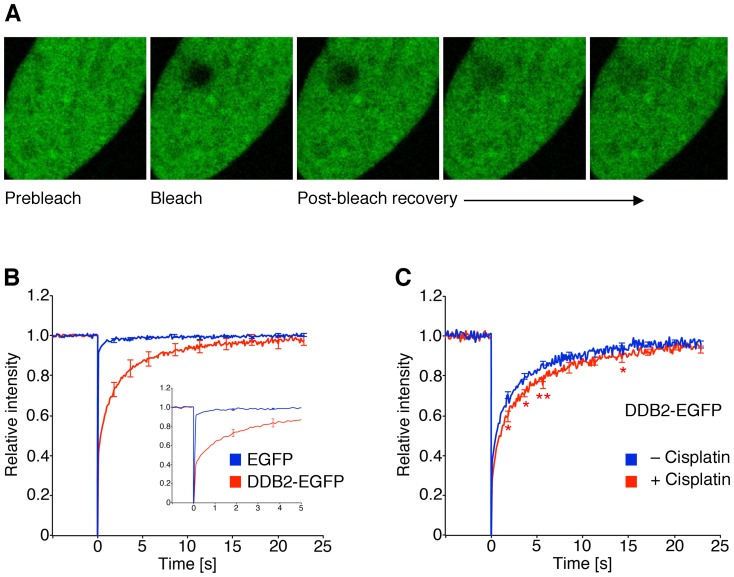
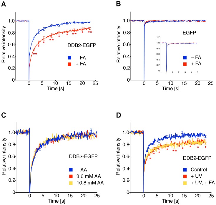
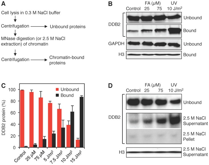
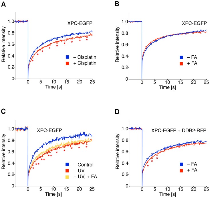
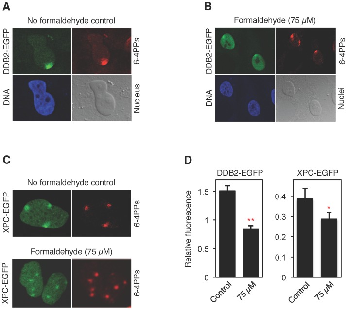
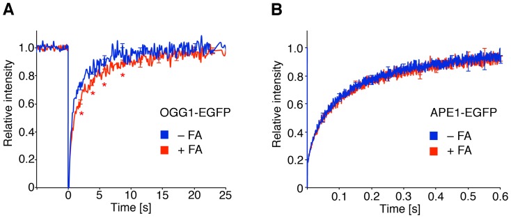
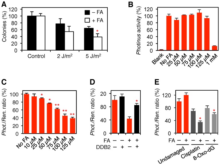
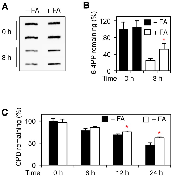
Methodology/principal findings: The overall mobility of the DNA damage sensors UV-DDB (ultraviolet-damaged DNA-binding) and XPC (xeroderma pigmentosum group C) was analyzed by assessing real-time protein dynamics in the nucleus of cultured human cells exposed to non-cytotoxic (<100 μM) formaldehyde concentrations. The DNA lesion-specific recruitment of these damage sensors was tested by monitoring their accumulation at local irradiation spots. DNA repair activity was determined in host-cell reactivation assays and, more directly, by measuring the excision of DNA lesions from chromosomes. Taken together, these assays demonstrated that formaldehyde obstructs the rapid nuclear trafficking of DNA damage sensors and, consequently, slows down their relocation to DNA damage sites thus delaying the excision repair of target lesions. A concentration-dependent effect relationship established a threshold concentration of as low as 25 micromolar for the inhibition of DNA excision repair.

Conclusions/significance: A main implication of the retarded repair activity is that low-dose formaldehyde may exert an adjuvant role in carcinogenesis by impeding the excision of multiple mutagenic base lesions. In view of this generally disruptive effect on DNA repair, we propose that formaldehyde exposures in the general population should be further decreased to help reducing cancer risks.


Conflict of interest statement

Competing Interests: The authors have declared that no competing interests exist.

Figures

Figure 1
Figure 1. Analysis of repair protein dynamics in living cells.
(A) A region of interest of ∼4 μm2 was photo-bleached and the fluorescence recovery within this area was monitored over a time frame of 23 seconds. Simultaneously, a reference area of the same size was monitored to correct for overall bleaching and the resulting data were normalized to the pre-bleach intensity. (B) Quantitative FRAP recordings determined in fibroblasts transfected with expression vectors coding for the DDB2-EGFP fusion or the EGFP moiety alone (N = 30; error bars, S.E.M.). (C) Recognition of cisplatin-DNA adducts by UV-DDB revealed by FRAP analyses. Human fibroblasts transfected with the DDB2-EGFP construct were pre-incubated with 5 μM cisplatin. The resulting FRAP curves were compared with those of untreated controls. Asterisks, statistically significant differences between cisplatin-treated cells and untreated controls (N = 50; *p<0.05; **p<0.01).
Figure 2
Figure 2. Delayed nuclear trafficking of UV-DDB.
(A) FRAP analysis in human fibroblasts. Cells were transfected with DDB2-EGFP, incubated for 18 h with 75 μM formaldehyde (FA) and analyzed by FRAP (N = 50; error bars, S.E.M.). The fluorescence recovery curves were compared to those of untreated controls (*p<0.05; **p<0.01). (B) FRAP studies (N = 15; error bars, S.E.M.) demonstrating that EGFP movements are not affected by formaldehyde (18 h, 75 μM). (C) FRAP analysis with analogous acetaldehyde (AA) treatments (N = 50). (D) Combined formaldehyde and UV treatment. Transfected human fibroblasts were exposed to 75 μM formaldehyde for 18 h, UV-irradiated (30 J/m2) and subjected to FRAP analysis (N = 30). The asterisks indicate significant differences between UV-damaged fibroblasts and untreated controls (*p<0.05; **p<0.01).
Figure 3
Figure 3. Association of UV-DDB with damaged chromatin.
(A) Flow diagram illustrating how chromatin was dissected to monitor the binding of UV-DDB. Unbound proteins were released by salt (0.3 M NaCl) extraction and the remaining chromatin was solubilized by MNase digestion. (B) Western blot visualization of the chromatin partitioning of UV-DDB using antibodies against DDB2. GAPDH (glyceraldehyde 3-phosphate dehydrogenase), marker of unbound proteins; histone H3, marker of chromatin. Human fibroblasts were exposed for 18 h to formaldehyde or UV-irradiated at the indicated doses. (C) Quantification of three independent binding assays demonstrating the differential interaction of DDB2 with formaldehyde- and UV-damaged chromatin (error bars, S.D.). (D) Release of chromatin-bound DDB2 and histone H3 by high-salt extraction. After incubation with 0.3 M NaCl buffer, the chromatin was dissolved with 2.5 M NaCl, thus liberating non-covalently bound chromatin proteins.
Figure 4
Figure 4. Indirect formaldehyde-induced reduction of XPC mobility.
(A) Protein dynamics studies of XPC in the nuclei of human fibroblasts. Cells were transfected with the XPC-EGFP construct, incubated with 5 μM cisplatin and subjected to FRAP analysis (N = 30; error bars, S.E.M.). The resulting fluorescence recovery curves were compared to those of untreated controls (*p<0.05). (B) FRAP studies (N = 50) demonstrating that the fluorescence recovery curves of XPC-EGFP are not affected by an 18-h formaldehyde treatment (75 μM). (C) FRAP analysis of transfected fibroblasts demonstrating that the 18-h formaldehyde treatment (75 μM) does not further reduce the delayed XPC-EGFP trafficking in UV-irradiated cells (30 J/m2; N = 50). (D) Combined formaldehyde treatment and DDB2 overexpression. The transfected fibroblasts were exposed to 75 μM formaldehyde for 18 h and subjected to FRAP analysis. The presence of DDB2-RFP resulted in a slightly delayed fluorescence recovery curve of XPC-EGFP upon formaldehyde exposure (N = 30). Asterisks, significant differences between formaldehyde-treated and untreated fibroblasts (*p<0.05).
Figure 5
Figure 5. Accumulation of damage recognition factors on UV lesions.
(A) Representative image illustrating the redistribution of DDB2 to UV lesion sites visualized with antibodies against (6-4) photoproducts. Human fibroblasts transfected with DDB2-EGFP were UV-irradiated through the pores of polycarbonate filters and fixed 15 min after treatment. DNA is evidenced by the Hoechst reagent and the nuclei are shown with contrast images. (B) Representative cells demonstrating the defective translocation of DDB2-EGFP from undamaged nuclear areas to UV lesions after 18-h incubations with 75 μM formaldehyde. (C) Representative images illustrating that the 75-μM formaldehyde treatment impedes the redistribution of XPC-EGFP to UV lesions. (D) Reduced fluorescence intensity at UV lesion spots over the surrounding background in cells exposed for 18 h to 75 μM formaldehyde (N = 54; error bars, S.D.; *p<0.05, **p<0.01).
Figure 6
Figure 6. Formaldehyde-induced damage delays the nuclear trafficking of a DNA glycosylase.
(A) Nuclear dynamics of OGG1, the DNA glycosylase that removes 8-oxo-dG, in human fibroblasts. Cells were transfected with the OGG1-EGFP construct, incubated for 18 h with 75 μM formaldehyde and subjected to FRAP analysis (N = 50; error bars, S.E.M.). The resulting fluorescence recovery curves were compared to those of untreated controls (*p<0.05). (B) FRAP studies (N = 50) demonstrating that the extremely fast movements of the APE1-EGFP fusion are not affected by the 75-μM formaldehyde treatment.
Figure 7
Figure 7. Inhibition of NER and BER activity by low-dose formaldehyde.
(A) Colony-forming assay demonstrating that human fibroblasts exposed to formaldehyde (75 μM) are more sensitive to killing by UV radiation (2 and 5 J/m2) than the respective untreated controls (error bars, S.D.; n = 3, each measurement in triplicate). The asterisk (*p<0.05) denotes the significantly reduced colony formation ability. (B) Expression of Photinus luciferase in cells containing undamaged pGL3 and exposed (18 h) to formaldehyde. All values (N = 3; error bars, S.D.) are shown as a percentage of luciferase activity in untreated fibroblasts. Blank, untransfected cells. (C) Host-cell reactivation of UV-irradiated pGL3 reflecting NER activity in cells exposed (18 h) to formaldehyde. Values (N = 10; error bars, S.D.) are shown as a percentage of the Photinus/Renilla ratio in control cells. Asterisks, significant differences from the control (*p<0.05, **p<0.01). (D) Partial restoration of host-cell reactivation in 100-μM formaldehyde-treated cells overexpressing DDB2-EGFP (N = 5). Asterisk, significantly (*p<0.05) higher NER activity then controls without DDB2-EGFP. (E) Inhibition of host-cell reactivation of pGL3 containing cisplatin adducts or 8-oxo-dG lesions (N = 5). The asterisks (*p<0.05) denote significantly reduced DNA repair activity in formaldehyde-treated (75 μM, 18 h) cells.
Figure 8
Figure 8. Inhibition of UV lesion removal from chromatin.
(A) Untreated or formaldehyde-treated cells (75 μM, 18 h) were exposed to UV light (10 J/m2) and collected immediately after irradiation or following 3-h repair incubations. Genomic DNA was isolated and analyzed for UV lesions using antibodies against 6-4PPs. (B) Quantification of three independent experiments demonstrating that 6-4PP excision is diminished by 75-μM formaldehyde exposure (error bar, S.D.). The asterisk (*p<0.05) denotes significantly reduced excision in formaldehyde-treated cells compared to the untreated control. (C) Quantification of three independent experiments demonstrating that CPD excision is also inhibited. The asterisks (*p<0.05) denote the significantly reduced excision in 75-μM formaldehyde-treated cells compared to untreated controls.

References

    1. Donaldson MR, Coldiron BM (2011) No end in sight: the skin cancer epidemic continues. Semin Cutan Med Surg 30: 3–5. - PubMed
    1. Hoeijmakers JH (2009) DNA damage, aging and cancer. New Engl J Med 361: 1475–1485. - PubMed
    1. Friedberg EC, Walker GC, Siede W, Wood RD, Schultz RA, et al. (2006) DNA Repair and Mutagenesis (ASM Press, Washington D.C).
    1. Parsons JL, Dianov GL (2013) Co-ordination of base excision repair and genome stability. DNA Rep 12: 326–333. - PubMed
    1. Maynard S, Schurmann SH, Harboe C, de Souza-Pinto NC, Bohr VA (2009) Base excision repair of oxidative DNA damage and association with cancer and aging. Carcinogenesis 30: 2–10. - PMC - PubMed

Publication types

LinkOut - more resources