. 2014 Jul;53(7):622-33.
doi: 10.1002/gcc.22172. Epub 2014 Apr 10.

Distinct transcriptional signature and immunoprofile of CIC-DUX4 fusion-positive round cell tumors compared to EWSR1-rearranged Ewing sarcomas: further evidence toward distinct pathologic entities

Affiliations

Distinct transcriptional signature and immunoprofile of CIC-DUX4 fusion-positive round cell tumors compared to EWSR1-rearranged Ewing sarcomas: further evidence toward distinct pathologic entities

Katja Specht et al. Genes Chromosomes Cancer. 2014 Jul.

Abstract

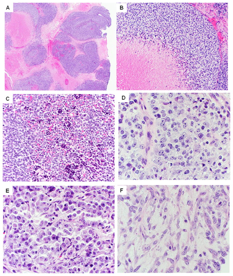
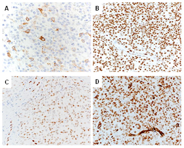
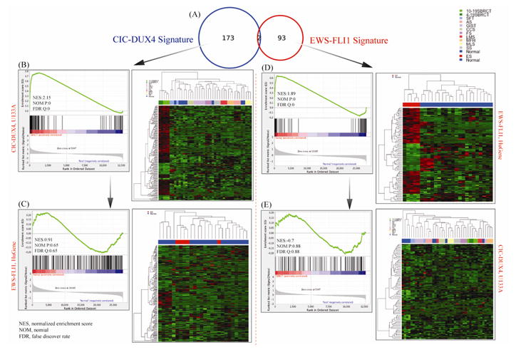
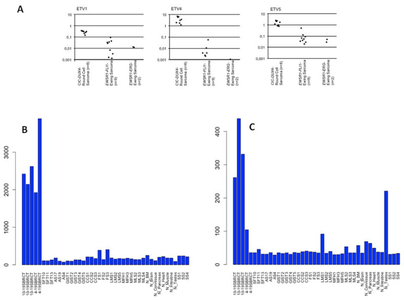
Round cell sarcomas harboring CIC-DUX4 fusions have recently been described as highly aggressive soft tissue tumors of children and young adults. Due to partial morphologic and immunohistochemical overlap with Ewing sarcoma (ES), CIC-DUX4-positive tumors have generally been classified as ES-like and managed similarly; however, a systematic comparison at the molecular and immunohistochemical levels between these two groups has not yet been conducted. Based on an initial observation that CIC-DUX4-positive tumors show nuclear immunoreactivity for WT1 and ETS transcription factors, FLI1 and ERG, we performed a detailed immunohistochemical and molecular analysis including these markers, to further investigate the relationship between CIC-DUX4 tumors and ES. The study group included 21 CIC-DUX4-positive sarcomas and 20 EWSR1-rearranged ES. Immunohistochemically, CIC-DUX4 sarcomas showed membranous CD99 positivity in 18 (86%) cases, but only 5 (24%) with a diffuse pattern, while WT1 and FLI1 were strongly positive in all cases. ERG was positive in 18% of cases. All ES expressed CD99 and FLI1, while ERG positivity was only seen in EWSR1-ERG fusion positive ES. WT1 was negative in all ES. Expression profiling validated by q-PCR revealed a distinct gene signature associated with CIC-DUX4 fusion, with upregulation of ETS transcription factors (ETV4, ETV1, and ETV5) and WT1, among top overexpressed genes compared to ES, other sarcomas and normal tissue. In conclusion, the distinct gene signature and immunoprofile of CIC-DUX4 sarcomas suggest a distinct pathogenesis from ES. The consistent WT1 expression may provide a useful clue in the diagnosis in the context of round cell sarcomas negative for EWSR1 rearrangement. © 2014 Wiley Periodicals, Inc.


Figures

Figure 1
Figure 1. Pathologic features of CIC-DUX4-positive sarcomas
A. At low-power, tumors show a vague nodular growth, often outlined by confluent geographic areas of necrosis (case 10) B. Higher power show tumor cells arranged in solid sheets, surrounding areas of necrosis (case 18). C. Admixed with viable cells are often pyknotic, degenerating cells (case 20; x100). D. Higher power showing ill-defined cell borders with vesicular chromatin and distinct nucleoli. Of note the nuclei show a round, oval to more angulated appearance, with subtle but increased pleomorphism than typical ES (case 16; x200). E. Case 10 displaying greater variability in nuclear size, prominent nucleoli and more abundant cytoplasm, pushing the nuclei at the periphery. F. Areas with spindle cell morphology are typically rare and present as a focal phenomenon. However, not uncommon is the presence of a edematous, myxoid stroma (case 16; 200x).
Figure 2
Figure 2. Immunohistochemical findings in CIC-DUX4 sarcoma
A. CD99 expression is typically focal (1+) and of moderate intensity (case 16). B. FLI1 labeling is uniformly strongly positive in the majority of tumor cells (case 16). C. ERG expression is variable, with multifocal (3+) and moderate staining intensity (case 10). D. WT-1 staining is seen in all CIC-DUX4 – positive sarcomas, with most cases displaying both cytoplasmic and nuclear staining (case 10).
Fig. 3
Fig. 3. Distinct Transcriptional Signature of CIC-DUX4-positive tumors compared to EWSR1-FLi1 Ewing sarcoma (ES) cases
A. Venn diagram showing minimal genomic overlap between the two groups investigated (n=2 genes). The 175 gene list was obtained by comparing 5 CIC-DUX4 tumors with 29 soft tissue sarcomas and 8 normal tissues on Affymetrix U133A chip (1.4 FC; FDR 0.05 p-value). The ES 95 gene-signature was obtained by overlapping the 854 differentially expressed genes in the 5 ES tumors (Affymetrix Hu-Gene; 1.3FC; FDR 0.1 p-value) with a published meta-analysis of ES. B. The 175 gene-signature was applied for hierarchical clustering showing a distinct genomic group of CIC-DUX4-tumors from all the other control samples. The GSEA confirms highly ranked genes, with high normalized enrichment score (NES) values. C. In contrast, applying the 175 CIC-DUX4 gene-signature to the ES and controls resulted in poor clustering expression patterns and low GSEA scores. D. Hierarchical clustering using the robust 95 ES gene-list shows a well-defined ES genomic cluster from all the normal controls, with high NES scores on GSEA. E. In contrast, using the same 95 gene-signature on the CIC-DUX4 tumors and controls shows an ambiguous expression pattern on clustering and low NEM score.
Fig. 4
Fig. 4
A. Quantitative RT-PCR reveals upregulation of ETV1, ETV4 and ETV5 in CIC-DUX4 sarcomas (n=8) as compared to EWSR1-FLI1 (n=9) or EWSR1-ERG (n=2) rearranged Ewing sarcomas (median ETV1: 24xfold; median ETV4: 1720x-fold; median ETV5: 30x-fold upregulation). B, C. Bar chart of mRNA overexpression of ETV4 (B) and WT1 (C) in CIC-DUX4-postiive tumors compared to other sarcoma types and normal tissue. WT1 shows overexpression mainly in the t(10;19) tumors.

References

    1. Amin KM, Litzky LA, Smythe WR, Mooney AM, Morris JM, Mews DJ, Pass HI, Kari C, Rodeck U, Rauscher FJ, Kaiser LR, Albelda SM. Wilms’ tumor 1 susceptibility (WT1) gene products are selectively expressed in malignant mesothelioma. Am J Pathol. 1995;146:344–356. - PMC - PubMed
    1. Antonescu CR, Viale A, Sarran L, Tschernyavsky SJ, Gonen M, Segal NH, Maki RG, Socci ND, DeMatteo RP, Besmer P. Gene expression in gastrointestinal stromal tumors is distinguished by KIT genotype and anatomic site. Clin Cancer Res. 2004;10:3282–3290. - PubMed
    1. Antonescu CR, Yoshida A, Guo T, Chang NE, Zhang L, Agaram NP, Qin LX, Brennan MF, Singer S, Maki RG. KDR activating mutations in human angiosarcomas are sensitive to specific kinase inhibitors. Cancer Res. 2009;69:7175–7179. - PMC - PubMed
    1. Barnoud R, Sabourin JC, Pasquier D, Ranchere D, Bailly C, Terrier-Lacombe MJ, Pasquier B. Immunohistochemical expression of WT1 by desmoplastic small round cell tumor: a comparative study with other small round cell tumors. Am J Surg Pathol. 2000;24:830–836. - PubMed
    1. Bettegowda C, Agrawal N, Jiao Y, Sausen M, Wood LD, Hruban RH, Rodriguez FJ, Cahill DP, McLendon R, Riggins G, Velculescu VE, Oba-Shinjo SM, Marie SK, Vogelstein B, Bigner D, Yan H, Papadopoulos N, Kinzler KW. Mutations in CIC and FUBP1 contribute to human oligodendroglioma. Science. 2011;333:1453–1455. - PMC - PubMed

Publication types

MeSH terms