. 2014 Jun;34(6):1162-70.
doi: 10.1161/ATVBAHA.114.302528. Epub 2014 Apr 10.

Lymphatic vessel insufficiency in hypercholesterolemic mice alters lipoprotein levels and promotes atherogenesis

Affiliations

Lymphatic vessel insufficiency in hypercholesterolemic mice alters lipoprotein levels and promotes atherogenesis

Taina Vuorio et al. Arterioscler Thromb Vasc Biol. 2014 Jun.

Abstract

Objective: Lymphatic vessels collect extravasated fluid and proteins from tissues to blood circulation as well as play an essential role in lipid metabolism by taking up intestinal chylomicrons. Previous studies have shown that impairment of lymphatic vessel function causes lymphedema and fat accumulation, but clear connections between arterial pathologies and lymphatic vessels have not been described.

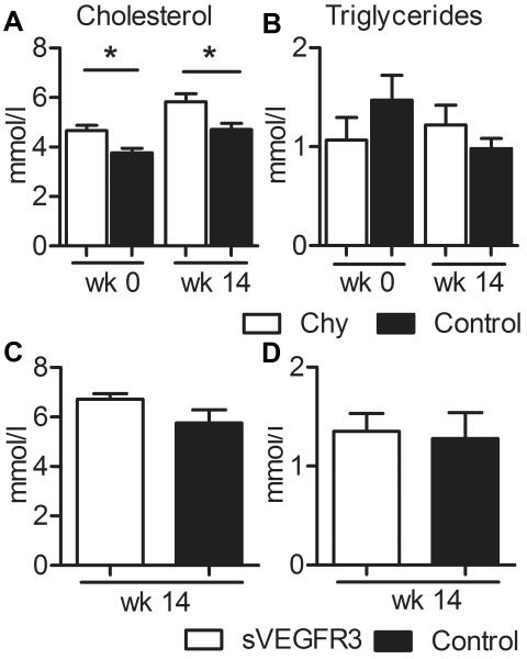
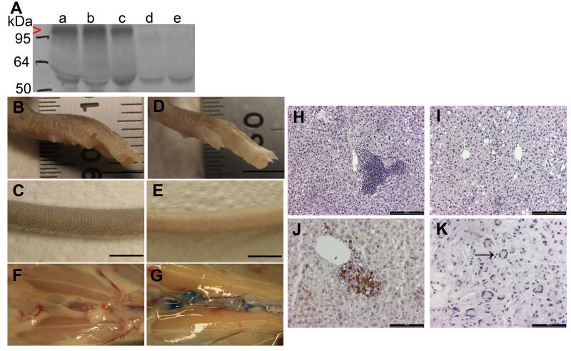
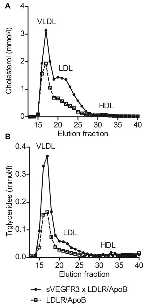
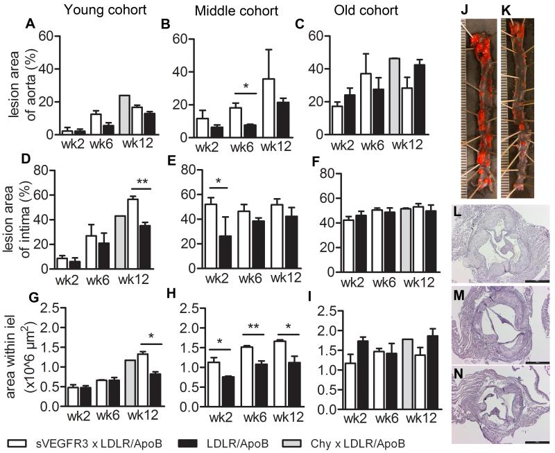
Approach and results: Two transgenic mouse strains with lymphatic insufficiency (soluble vascular endothelial growth factor 3 [sVEGFR3] and Chy) were crossed with atherosclerotic mice deficient of low-density lipoprotein receptor and apolipoprotein B48 (LDLR(-/-)/ApoB(100/100)) to study the effects of insufficient lymphatic vessel transport on lipoprotein metabolism and atherosclerosis. Both sVEGFR3×LDLR(-/-)/ApoB(100/100) mice and Chy×LDLR(-/-)/ApoB(100/100) mice had higher plasma cholesterol levels compared with LDLR(-/-)/ApoB(100/100) control mice during both normal chow diet (16.3 and 13.7 versus 8.2 mmol/L, respectively) and Western-type high-fat diet (eg, after 2 weeks of fat diet, 45.9 and 42.6 versus 30.2 mmol/L, respectively). Cholesterol and triglyceride levels in very-low-density lipoprotein and low-density lipoprotein fractions were increased. Atherosclerotic lesions in young and intermediate cohorts of sVEGFR3×LDLR(-/-)/ApoB(100/100) mice progressed faster than in control mice (eg, intermediate cohort mice at 6 weeks, 18.3% versus 7.7% of the whole aorta, respectively). In addition, lesions in sVEGFR3×LDLR(-/-)/ApoB(100/100) mice and Chy×LDLR(-/-)/ApoB(100/100) mice had much less lymphatic vessels than lesions in control mice (0.33% and 1.07% versus 7.45% of podoplanin-positive vessels, respectively).

Conclusions: We show a novel finding linking impaired lymphatic vessels to lipoprotein metabolism, increased plasma cholesterol levels, and enhanced atherogenesis.

Keywords: atherosclerosis; lipoproteins; lymphatic vessels.


Figures

Figure 1
Figure 1
Effects of Western diet on Chy and sVEGFR3 mice. Total cholesterol (A) and triglycerides (B) were measured in male Chy mice and their wild type littermates on chow diet and after 14 wk on Western diet and in male sVEGFR3 mice and their littermates after 14 wk on Western diet (C and D, respectively). Conversion factors from mmol/l to mg/dl for cholesterol and triglycerides are 38.67 and 88.57, respectively. Data is presented as mean ± SEM. * p < 0.05.
Figure 2
Figure 2
Characterization of the sVEGFR3 × LDLR−/−/ApoB100/100 mouse model. Expression of sVEGFR3 in plasma was confirmed by western blotting (A). sVEGFR3 was expressed in sVEGFR3 × LDLR−/−/ApoB100/100 mice (lanes a, b and c, red arrowhead), but not in LDLR−/−/ApoB100/100 mice (lanes d and e). Antibody cross-reacted with mouse IgG heavy chain (55 kDa). Lymphatic drainage was affected in sVEGFR3 × LDLR−/−/ApoB100/100 mice causing swelling in the feet (B) and tails (C) compared to LDLR−/−/ApoB100/100 mice (D and E, respectively). Lymphatic insufficiency was confirmed with Evans Blue dye injections into footpads. No visible dye was detected in the abdominal collecting lymphatic vessels in sVEGFR3 × LDLR−/−/ApoB100/100 mice 30 min after dye injection (F), whereas collecting lymphatic vessels in LDLR−/−/ApoB100/100 mice were clearly stained (G). On Western diet, sVEGFR3 × LDLR−/−/ApoB100/100 mice had accumulation of inflammatory cells around hepatic central veins in the liver (H) whereas livers in LDLR−/−/ApoB100/100 mice were normal (I). Most inflammatory cells were CD3 positive T-lymphocytes (J). Compromised lymphatic function caused also swelling in the jaw area of sVEGFR3 × LDLR−/−/ApoB100/100 mice characterized by cholesterol crystal-like structures and multinuclear inflammatory giant cells (K, black arrow). Bar 5 mm (B-E), 200 μm (H and I) and 100 μm (J and K).
Figure 3
Figure 3
Analysis of cholesterol and triglyceride levels in plasma lipoproteins. Distribution of cholesterol (A) and triglycerides (B) in different lipoprotein fractions in young sVEGFR3 × LDLR−/−/ApoB100/100 mice fed Western diet for 6 wk show clear increases in VLDL and LDL fractions in sVEGFR3 × LDLR−/−/ApoB100/100 mice compared to LDLR−/−/ApoB100/100 mice. Both genders have been used in the analysis.
Figure 4
Figure 4
Evaluation of atherosclerotic lesion areas in sVEGFR3 × LDLR−/−/ApoB100/100 mice, Chy × LDLR−/−/ApoB100/100 mice and LDLR−/−/ApoB100/100 mice. Data is presented as the total en face lesion area of the total aortic area (A, B and C), as the total intimal lesion area of the aortic root (D, E and F) and as area within internal elastic lamina (G, H and I). Lesion areas were measured on 2, 6 and 12 weeks after starting the Western diet in the young, intermediate and old cohorts (3-4, 7-8, and 11-12 months of age, respectively). Both genders were used in the analysis. Representative pictures of En face aortas in intermediate cohort of sVEGFR3 × LDLR−/−/ApoB100/100 mice (J) and LDLR−/−/ApoB100/100 mice (K) after 4 weeks of Western diet and Hematoxylin-Eosin stainings showing atherosclerotic lesions in young cohorts sVEGFR3 × LDLR−/−/ApoB100/100 mice (G), Chy × LDLR−/−/ApoB100/100 mice (H) and LDLR−/−/ApoB100/100 mice (I) after 12 weeks of Western diet. Data is presented as mean ± SEM. * p < 0.05, ** p < 0.01. Unit 500 μm (J and K) and bar 500 μm (L-N).
Figure 5
Figure 5
Evaluation of lesion composition in sVEGFR3 × LDLR−/−/ApoB100/100 mice, Chy × LDLR−/−/ApoB100/100 mice and LDLR−/−/ApoB100/100 mice. Representative pictures of modified Movat’s pentachrome stainings of atherosclerotic lesions in aortic roots after 2, 6 and 12 weeks of Western diet. Intermediate cohort sVEGFR3 × LDLR−/−/ApoB100/100 mice show increased amounts of cholesterol crystals (arrow) and collagen compared to LDLR−/−/ApoB100/100 control mice (A-F). mMQ stainings for macrophages in intermediate cohort sVEGFR3 × LDLR−/−/ApoB100/100 mice (G) and LDLR−/−/ApoB100/100 mice (H) after 6 weeks of fat diet show accumulation of macrophages and expanding core in a lesion in sVEGFR3 × LDLR−/−/ApoB100/100 mice. No difference in lipid deposition was detected between strains in Oil-red-O for intermediate after 4 weeks of Western diet (I and J). Movat’s staining (K) and mMQ (L) staining for lesions in young cohort Chy × LDLR−/−/ApoB100/100 mice after 12 weeks of Western diet show accumulation of cholesterol crystals and some macrophages. Bar 200 μm.
Figure 6
Figure 6
Confocal images of neovascularization and lymphatic vessels associated with atherosclerotic lesions in descending aortas of old cohort sVEGFR3 × LDLR−/−/ApoB100/100 mice (A), Chy × LDLR−/−/ApoB100/100 mice (B) and LDLR−/−/ApoB100/100 mice (C) on Western diet. See Methods for in vivo and whole mount in situ staining. Images shown are Z-stack projections of vessels in depth of 24 μm from the adventitia into the media. Intravascularly labeled GSL-I lectin vessels (red) colocalized with most but not all CD31-reactive vessels (green). Podoplanin-positive vessels (magenta) had a larger caliber and expressed CD31 but did not contain GSL-I lectin. The densities of vasa vasorum in plaques (D) (open bars) from all groups were similar (sVEGFR3 × LDLR−/−/ApoB100/100 median 11.2 % area of GSLI/hpf range 4.36-21.34, Chy × , LDLR−/−/ApoB100/100 median 9.18 %, range 4.03-17.2 and LDLR−/−/ApoB100/100 controls median 8.20%, range 2.45-17. (Kruskal Wallis P = 0.5491, Dunn’s post test comparison between groups P > 0.05)). Lymphatic vessels (grey bars) were nearly absent or irregularly formed in sVEGFR3 × LDLR−/−/ApoB100/100 mice (median 0.33% area podoplanin+ vessels / hpf, range 0.09-1.33) and Chy × LDLR−/−/ApoB100/100 mice (median 1.07%, range 0.04-4.36) compared to LDLR−/−/ApoB100/100 controls (median 7.45%, range 0.12-19.59) (Kruskal Wallis P = 0.0014, Dunn’s post test comparisons P < 0.05 for sVEGFR3 ** and Chy* relative to controls). Both genders were used in the analysis. Single slice images of lymphatic vessels located at 8 μm depth in the adventitia (E) show spatial localization of podoplanin staining (magneta) with some CD31+ (green) vessels.

References

    1. Glass CK, Witztum JL. Atherosclerosis. the road ahead. Cell. 2001;104:503–516. - PubMed
    1. Lusis AJ. Atherosclerosis. Nature. 2000;407:233–241. - PMC - PubMed
    1. Ross R. The pathogenesis of atherosclerosis: a perspective for the 1990s. Nature. 1993;362:801–809. - PubMed
    1. Moulton KS. Angiogenesis in atherosclerosis: gathering evidence beyond speculation. Curr Opin Lipidol. 2006;17:548–555. - PubMed
    1. Yla-Herttuala S, Bentzon JF, Daemen M, et al. Stabilisation of atherosclerotic plaques. Position paper of the European Society of Cardiology (ESC) Working Group on atherosclerosis and vascular biology. Thromb Haemost. 2011;106:1–19. - PubMed

Publication types