. 2014 May;15(5):548-56.
doi: 10.1002/embr.201337631. Epub 2014 Apr 10.

PICK1 links Argonaute 2 to endosomes in neuronal dendrites and regulates miRNA activity

Affiliations

PICK1 links Argonaute 2 to endosomes in neuronal dendrites and regulates miRNA activity

Anna Antoniou et al. EMBO Rep. 2014 May.

Abstract

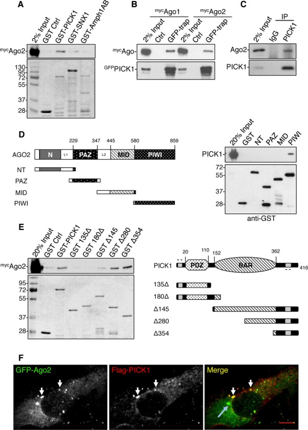
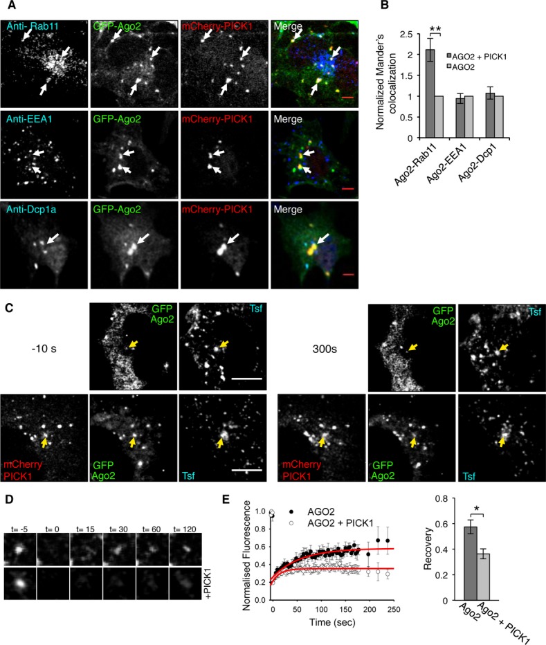
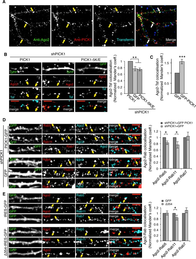
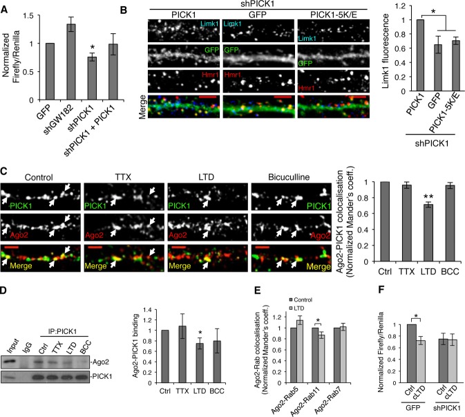
MicroRNAs fine-tune gene expression by inhibiting the translation of mRNA targets. Argonaute (Ago) proteins are critical mediators of microRNA-induced post-transcriptional silencing and have been shown to associate with endosomal compartments, but the molecular mechanisms that underlie this process are unclear, especially in neurons. Here, we report a novel interaction between Ago2 and the BAR-domain protein, PICK1. We show that PICK1 promotes Ago2 localization at endosomal compartments in neuronal dendrites and inhibits Ago2 function in translational repression following neuronal stimulation. We propose that PICK1 provides a link between activity-dependent endosomal trafficking and local regulation of translation in neurons.


Figures

Figure 1
Figure 1. PICK1 interacts with Ago2
A Ago2 binds to GST-PICK1 but not other BAR-domain proteins tested. GST-PICK1, GST-SNX1 (sorting nexin 1), and GST-Amph1AB (Amphiphysin1 domains AB) were incubated with HEK293 cell lysate expressing mycAgo2. Bound proteins were detected by western blotting using anti-myc or by Coomassie staining. B GFP-PICK1 interacts with both mycAgo1 and mycAgo2 from HEK293 cells. Lysates were incubated with GFP-trap agarose or blocked agarose beads as control, and bound proteins were detected by western blotting using anti-myc or anti-GFP. C Ago2 interacts with PICK1 in neurons. Lysates were immunoprecipitated with anti-PICK1 or control IgG, and bound proteins were detected by western blotting. D PICK1 interacts with the PIWI domain of Ago2. GST fusions of truncation mutants for the N-terminus (GST-NT) or the PAZ, MID, or PIWI domains as depicted in the diagram were incubated with purified his6-PICK1. Bound protein was detected by western blotting. E Ago2 interacts with the C-terminus of PICK1. GST-PICK1 full-length or truncation mutants as depicted in the diagram were incubated with HEK293 cell lysate expressing mycAgo2. Bound proteins were detected by western blotting with anti-myc or by Coomassie staining. F PICK1 colocalizes with Ago2 puncta distinct from P-bodies. COS7 cells expressing GFP-Ago2 and flag-PICK1 were stained with anti-flag. White arrows indicate overlapping puncta between Ago2 and PICK1; blue arrow indicates a P-body; scale bar, 10 μm.
Figure 2
Figure 2. PICK1 promotes Ago2 association with recycling endosomes
A PICK1 and Ago2 colocalize in Rab11-positive endosomal compartments in COS7 cells. Cells expressing GFP-Ago2 and mCherry-PICK1 were stained using specific antibodies against Rab11, EEA1, or Dcp1a (blue channel). Arrows indicate Ago2 and PICK1 overlapping puncta; scale bar, 5 μm. B PICK1 co-expression causes an increase in colocalization between Ago2 and Rab11. Colocalization analysis between Ago2 and Rab11, EEA1 or Dcp1a, was performed in COS7 cells expressing either GFP-Ago2 and mCherry-PICK1 or GFP-Ago2 alone. Graph shows Mander’s colocalization coefficients for the fraction of Ago2 colocalized with Rab11, EEA1, or Dcp1a in cells expressing both GFP-Ago2 and mCherry-PICK1 normalized to cells expressing Ago2 alone. **P = 0.0002 (Student’s t-test), n > 30 cells per condition. C Fluorescence recovery after photobleaching (FRAP) of GFP-Ago2 puncta colocalizing with transferrin-Alexa 647 (Tfn) in the absence or presence of mCherry-PICK1 in COS7 cells; scale bar, 10 μm. Images show cells pre-bleach (−10 s, left panels) and post-recovery (300 s, right panels). Yellow arrows indicate analyzed puncta. D Representative images of zoomed GFP-Ago2. Time (t) is in seconds; bleaching at t = 0. E Quantification of FRAP in (C) shows a decrease in recovery of Ago2 in the presence of PICK1. Fluorescence intensity was normalized to pre-bleach values, and fitted curves were used to extract recovery values. *P = 0.04 (Student’s t-test), n = 6–7 cells per condition.
Figure 3
Figure 3. PICK1 promotes Ago2 localization at endosomal compartments in dendrites of hippocampal neurons
A Endogenous Ago2 and PICK1 are found at transferrin-positive compartments in neuronal dendrites. Neurons were incubated with Alexa-conjugated transferrin to label endosomes and stained with Ago2 and PICK1 antibodies. Arrows indicate colocalizing puncta; scale bar, 10 μm. B PICK1 knockdown reduces Ago2 colocalization with endosomes. Neurons expressing shPICK1 plus GFP, sh-resistant GFP-PICK WT, or sh-resistant GFP-PICK 5K/E mutant were incubated with Alexa-conjugated transferrin (Tfn) and stained for Ago2. Bottom panels show the merge of Ago2 and Tfn channels. Scale bars, 5 μm. Graph shows Mander’s coefficients for the fraction of Ago2 colocalized with Tfn, normalized to GFP-PICK1 WT rescue condition. Arrows indicate overlapping puncta positive for Ago2 and transferrin. **P < 0.005 (Student’s t-test with Bonferroni correction), n > 10 cells per condition. C PICK1 overexpression increases Ago2 colocalization with endosomes. Cells expressing GFP or GFP-PICK1 were processed and analyzed as in (B). Representative images are shown in Supplementary Fig S2A. Graph shows Mander’s colocalization coefficients for the fraction of Ago2 colocalized with Tfn, normalized to GFP condition. ***P < 0.001, n > 10 cells per condition. D PICK1 knockdown reduces Ago2 colocalization with Rab5 and Rab11. Neurons expressing shPICK1 plus GFP or sh-resistant GFP-PICK1 were stained for Ago2 (cyan) and Rab5, Rab11, or Rab7 (red). Far right panels show the merge of Ago2 and Rab channels. Arrows indicate overlapping puncta positive for Ago2 and Rab protein; scale bars, 5 μm. Graph shows Mander’s coefficients for the fraction of Ago2 colocalized with Rab protein, normalized to GFP-PICK1 wild-type rescue condition. n > 8 cells per condition, *P < 0.05 (Student’s t-test), **P < 0.01. E Δ354 expression reduces Ago2 colocalization with Rab11. Neurons expressing empty-IRES-GFP or Δ354-IRES-EGFP were processed as in (D). Arrows indicate overlapping puncta positive for Ago2 and Rab protein. Graph shows Mander’s coefficients for the fraction of Ago2 colocalized with Rab protein, normalized to GFP control. *P = 0.03 (Student’s t-test), n > 9 cells per condition. Source data are available online for this figure.
Figure 4
Figure 4. PICK1 is involved in microRNA-mediated silencing pathways during chemical LTD
A PICK1 knockdown causes an increase in translational repression in cultured neurons. Dual-luciferase assay was performed in neurons expressing Renilla and Firefly luciferase reporter containing Limk1 3′UTR and either GFP, shGW182, shPICK1, or shPICK1 + sh-resistant GFP-PICK1. Values were normalized to the GFP-alone condition. *P < 0.05 (Student’s t-test with Bonferroni correction), n = 4–7. B PICK1 knockdown causes a decrease in endogenous Limk1 expression in neuronal dendrites. Neurons expressing shPICK1 plus GFP, sh-resistant GFP-PICK wild-type, or sh-resistant GFP-PICK 5K/E mutant were stained for Limk1 (blue) and the synaptic marker Homer1 (red). Scale bars, 5 μm. Graph shows Limk1 immunofluorescence normalized to GFP-PICK1 wild-type rescue condition. *P < 0.05 (Student’s t-test with Bonferroni correction), n > 14 cells per condition. C Chemical LTD causes a decrease in colocalization between Ago2 and PICK1 in dendrites of hippocampal neurons. Hippocampal neurons were treated with tetrodotoxin (TTX), Bicuculline (BCC), or NMDA (LTD), as shown and stained for PICK1 (green) and Ago2 (red). Arrows indicate overlapping puncta positive for PICK1 and Ago2. Scale bars, 5 μm. Graph shows Mander’s coefficients for the fraction of Ago2 colocalized with PICK1, normalized to untreated controls. **P < 0.01 (Student’s t-test with Bonferroni correction), n > 16 cells per condition. D Chemical LTD reduces the Ago2–PICK1 interaction. Neuronal cultures were treated as in (C), lysates were immunoprecipitated with anti-PICK1 or control IgG and bound proteins detected by western blotting. Values for Ago2 were normalized to PICK1 IP and to untreated control. *P < 0.05 (Student’s t-test with Bonferroni correction), n = 5–7. E Chemical LTD reduces the colocalization between Ago2 and Rab11. Neurons were treated for chemical LTD and stained with Ago2 and Rab proteins. Representative images are shown in Supplementary Fig S2B. Graph shows Mander’s coefficients for the fraction of Ago2 colocalized with Rab protein, normalized to untreated controls. *P = 0.03 (Student’s t-test), n > 9 cells per condition. F PICK1 knockdown occludes the effect of chemical LTD on translational repression. Dual-luciferase assay was performed in cultured neurons expressing GFP or GFP + shPICK1 and luciferase constructs as in (C). Neurons were treated for chemical LTD and lysed after 5 min. Relative luciferase values were normalized to vehicle-treated GFP control. *P = 0.03 (Student’s t-test), n = 8. Source data are available online for this figure.

References

    1. Kim VN, Han J, Siomi MC. Biogenesis of small RNAs in animals. Nat Rev Mol Cell Biol. 2009;10:126–139. - PubMed
    1. Djuranovic S, Nahvi A, Green R. A parsimonious model for gene regulation by miRNAs. Science. 2011;331:550–553. - PMC - PubMed
    1. McNeill E, Van Vactor D. MicroRNAs shape the neuronal landscape. Neuron. 2012;75:363–379. - PMC - PubMed
    1. Im HI, Kenny PJ. MicroRNAs in neuronal function and dysfunction. Trends Neurosci. 2012;35:325–334. - PMC - PubMed
    1. Siegel G, Saba R, Schratt G. microRNAs in neurons: manifold regulatory roles at the synapse. Curr Opin Genet Dev. 2011;21:491–497. - PubMed

MeSH terms

LinkOut - more resources