. 2012 Dec 1:188:312-319.
doi: 10.1016/j.sna.2011.12.044.

Light-Directed Migration of D. discoideum Slugs in Microfabricated Confinements

Affiliations

Light-Directed Migration of D. discoideum Slugs in Microfabricated Confinements

Jinho Kim et al. Sens Actuators A Phys. .

Abstract

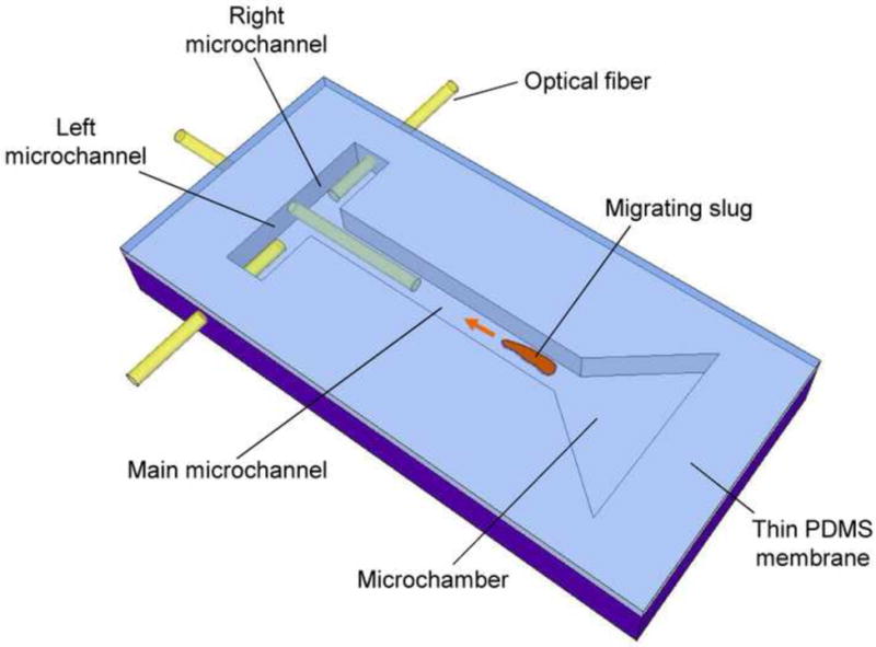
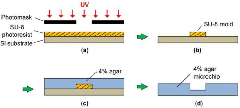
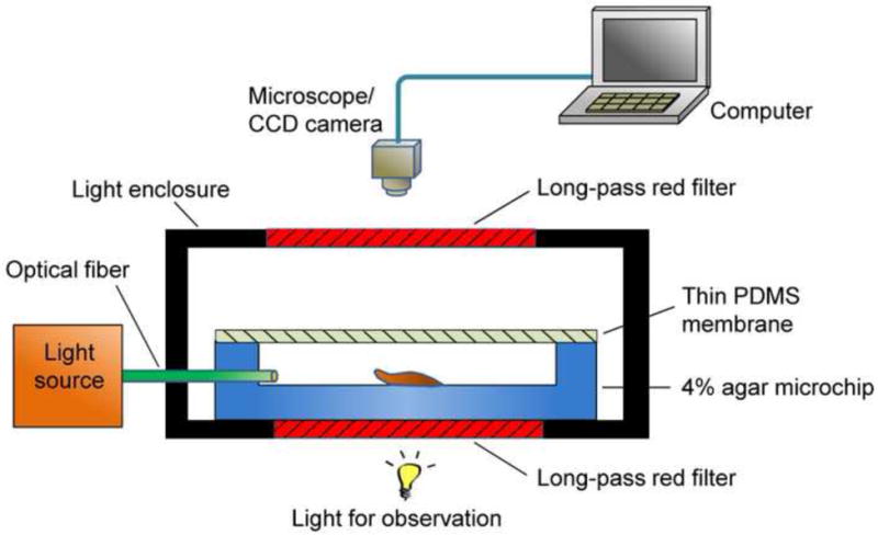
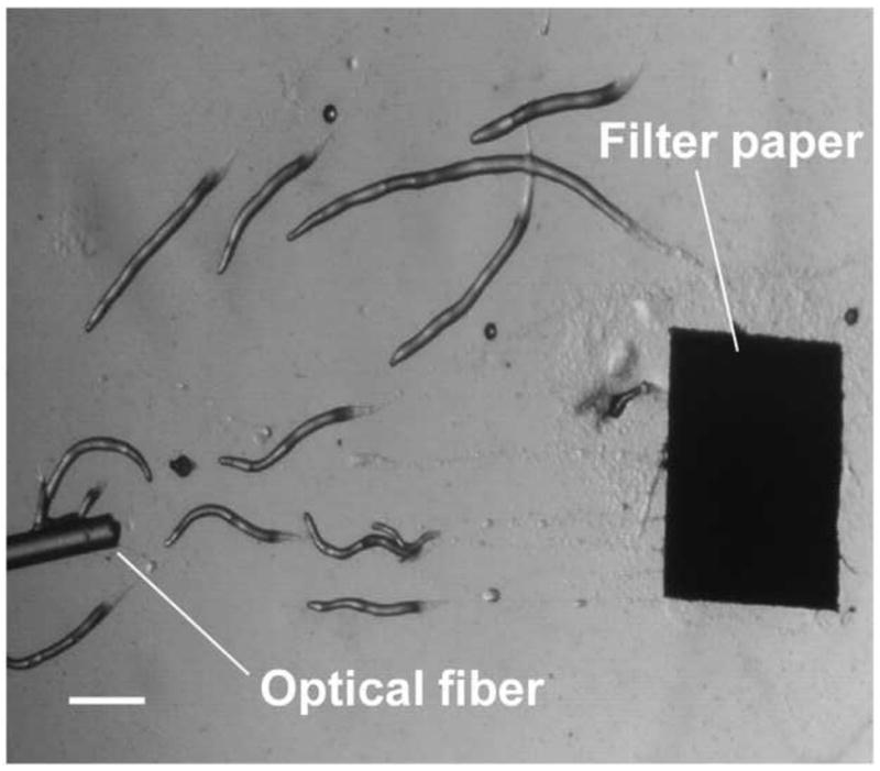
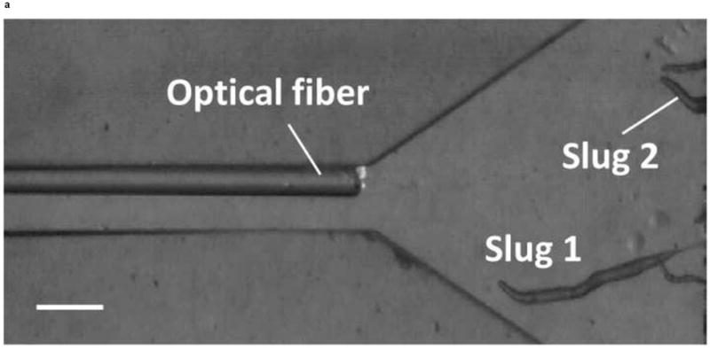
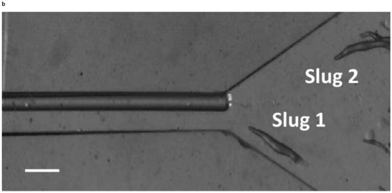
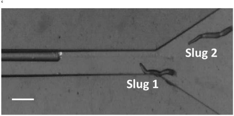
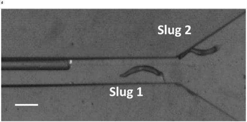
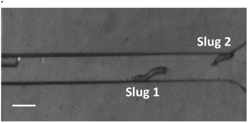
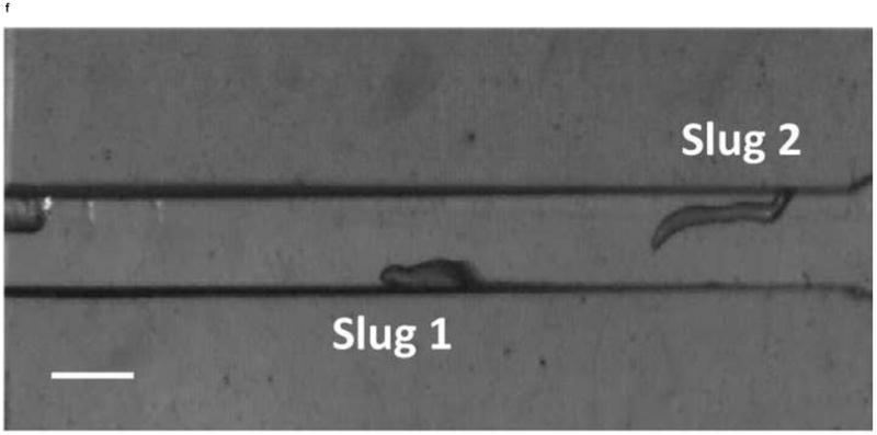
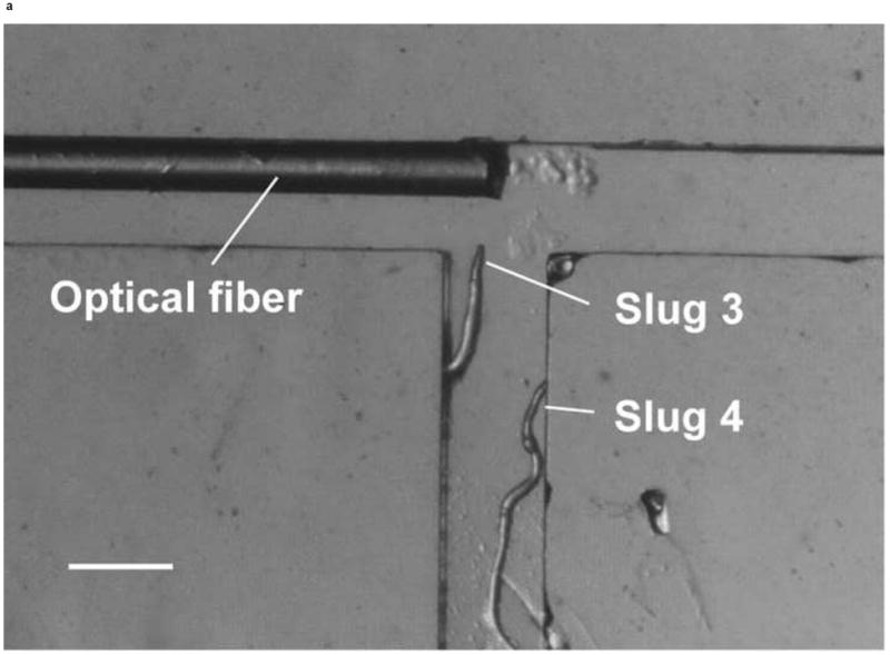
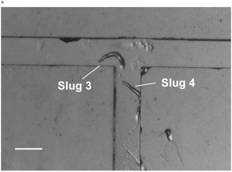
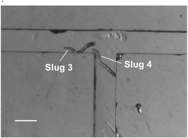
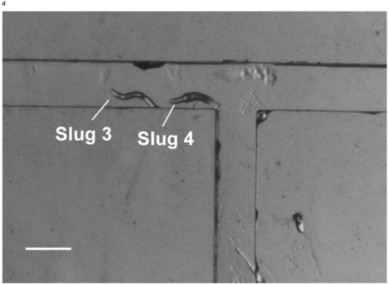
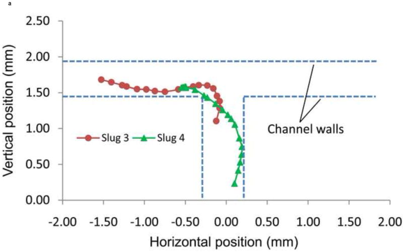
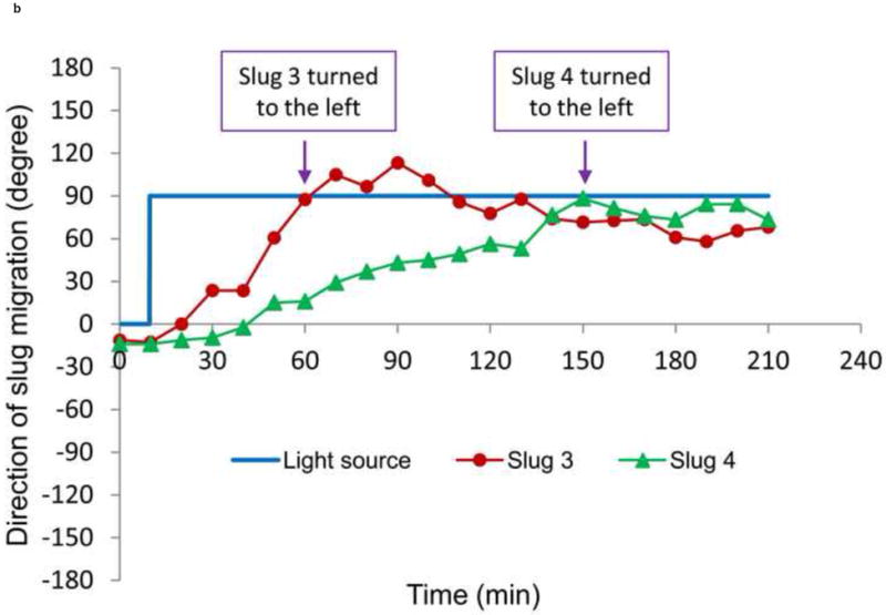
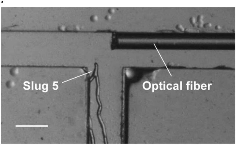
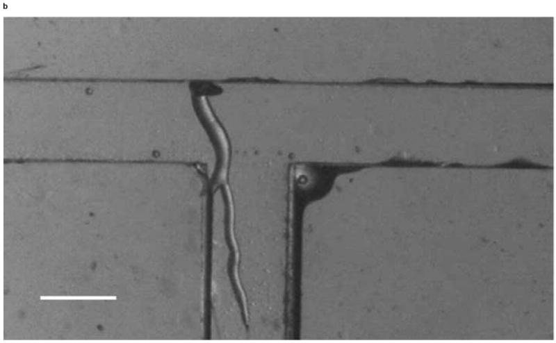
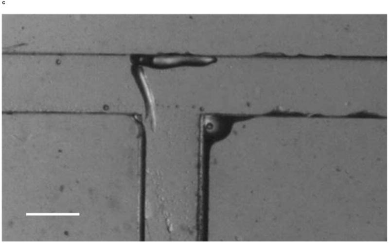
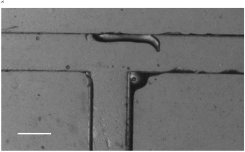
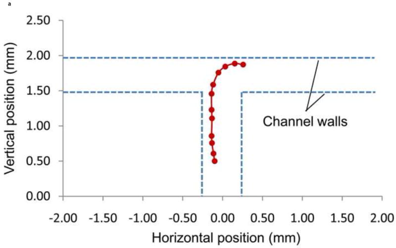
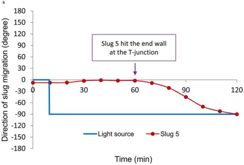
This paper investigates the light-driven migration of the multi-cellular microorganism Dictyostelium discoideum as a potential bio-actuation mechanism in microsystems. As a platform for slug migration we use microscale confinements, which consist of intersecting microchannels fabricated from solidified agar-water solution. The agar surface provides necessary moisture to the slugs during the experiment while remaining sufficiently stiff to allow effective slug migration. The movements of the slugs in the microchannels are driven and guided by phototaxis via controlling light transmitted through optical fibers. The microchannels impose geometrical confinements on the migrating slugs, improving the spatial precision of the migration. We demonstrate that slugs that form in a microchamber can be driven to migrate through the microchannels, as well as steered to a particular direction at microchannel intersections. Our experimental results indicate that slug movements can be more effectively controlled in microchannels, and potentially useful for bio-actuation applications.

Keywords: Bio-microactuator; Dictyostelium discoideum; microchannel; microorganism; phototaxis.


Figures

Figure 1
Figure 1
A schematic of phototaxis of a D. discoideum slug in a microchip. Migration of the slug is driven and directed by light transmitted through the optical fibers.
Figure 2
Figure 2
An illustration of the molding procedure of 4% agar microchannels. (a) Spin coated SU-8 photoresist (thickness: 500 μm) is exposed to UV light through a photomask. (b) Development produces a SU-8 mold. (c) Four percent molten agar is pour over the SU-8 mold and cured at room temperature for 10-15 minutes. (d) The solidified 4% agar microchip is then peeled off from the mold.
Figure 3
Figure 3
A schematic of a light enclosure for phototaxis experiment of slugs.
Figure 4
Figure 4
A micrograph image of slugs migrating toward an optical fiber light source on an open 4% agar surface. (Scale bar: 500 μm.)
Figure 5
Figure 5
Micrograph images of Slugs 1 and 2 at different times as the slugs migrated from the microchamber into and through the main microchannel: (a) t = 0 hr, (b) t = 1 hr, (c) t = 2 hr, (d) t = 3 hr, (e) t = 4 hr, and (f) t = 5 hr (Scale bar: 500 μm.)
Figure 5
Figure 5
Micrograph images of Slugs 1 and 2 at different times as the slugs migrated from the microchamber into and through the main microchannel: (a) t = 0 hr, (b) t = 1 hr, (c) t = 2 hr, (d) t = 3 hr, (e) t = 4 hr, and (f) t = 5 hr (Scale bar: 500 μm.)
Figure 5
Figure 5
Micrograph images of Slugs 1 and 2 at different times as the slugs migrated from the microchamber into and through the main microchannel: (a) t = 0 hr, (b) t = 1 hr, (c) t = 2 hr, (d) t = 3 hr, (e) t = 4 hr, and (f) t = 5 hr (Scale bar: 500 μm.)
Figure 5
Figure 5
Micrograph images of Slugs 1 and 2 at different times as the slugs migrated from the microchamber into and through the main microchannel: (a) t = 0 hr, (b) t = 1 hr, (c) t = 2 hr, (d) t = 3 hr, (e) t = 4 hr, and (f) t = 5 hr (Scale bar: 500 μm.)
Figure 5
Figure 5
Micrograph images of Slugs 1 and 2 at different times as the slugs migrated from the microchamber into and through the main microchannel: (a) t = 0 hr, (b) t = 1 hr, (c) t = 2 hr, (d) t = 3 hr, (e) t = 4 hr, and (f) t = 5 hr (Scale bar: 500 μm.)
Figure 5
Figure 5
Micrograph images of Slugs 1 and 2 at different times as the slugs migrated from the microchamber into and through the main microchannel: (a) t = 0 hr, (b) t = 1 hr, (c) t = 2 hr, (d) t = 3 hr, (e) t = 4 hr, and (f) t = 5 hr (Scale bar: 500 μm.)
Figure 6
Figure 6
Migration distances of Slugs 1 and 2 in the main microchannel.
Figure 7
Figure 7
Average speeds of slugs in microchannels as a function of slug length.
Figure 8
Figure 8
Micrograph images of Slugs 3 and 4 at different times as the slugs turned from the main channel into the left channel: (a) t = 0 min, (b) t = 60 min, (c) t = 120 min, and (d) t = 180 min (Scale bar: 500 μm.)
Figure 8
Figure 8
Micrograph images of Slugs 3 and 4 at different times as the slugs turned from the main channel into the left channel: (a) t = 0 min, (b) t = 60 min, (c) t = 120 min, and (d) t = 180 min (Scale bar: 500 μm.)
Figure 8
Figure 8
Micrograph images of Slugs 3 and 4 at different times as the slugs turned from the main channel into the left channel: (a) t = 0 min, (b) t = 60 min, (c) t = 120 min, and (d) t = 180 min (Scale bar: 500 μm.)
Figure 8
Figure 8
Micrograph images of Slugs 3 and 4 at different times as the slugs turned from the main channel into the left channel: (a) t = 0 min, (b) t = 60 min, (c) t = 120 min, and (d) t = 180 min (Scale bar: 500 μm.)
Figure 9
Figure 9
(a) Trajectory, and (b) direction of migration of Slugs 3 and 4 as it was turning to the left during phototaxis.
Figure 9
Figure 9
(a) Trajectory, and (b) direction of migration of Slugs 3 and 4 as it was turning to the left during phototaxis.
Figure 10
Figure 10
Micrograph images of Slug 5 at different times as the slugs turned from the main channel into the right channel: (a) t = 0 min, (b) t = 60 min, (c) t = 90 min, and (d) t = 120 min (Scale bar: 500 μm.)
Figure 10
Figure 10
Micrograph images of Slug 5 at different times as the slugs turned from the main channel into the right channel: (a) t = 0 min, (b) t = 60 min, (c) t = 90 min, and (d) t = 120 min (Scale bar: 500 μm.)
Figure 10
Figure 10
Micrograph images of Slug 5 at different times as the slugs turned from the main channel into the right channel: (a) t = 0 min, (b) t = 60 min, (c) t = 90 min, and (d) t = 120 min (Scale bar: 500 μm.)
Figure 10
Figure 10
Micrograph images of Slug 5 at different times as the slugs turned from the main channel into the right channel: (a) t = 0 min, (b) t = 60 min, (c) t = 90 min, and (d) t = 120 min (Scale bar: 500 μm.)
Figure 11
Figure 11
(a) Trajectory, and (b) direction of migration of Slug 5 as it was turning to the right during phototaxis.
Figure 11
Figure 11
(a) Trajectory, and (b) direction of migration of Slug 5 as it was turning to the right during phototaxis.

Similar articles

Cited by

References

    1. Weibel D, DiLuzio W, Whitesides G. Microfabrication meets microbiology. Nature Reviews Microbiology. 2007;5:209–218. - PubMed
    1. Steager E, Kim C, Patel J, Bith S, Naik C, Reber L, Kim M. Control of microfabricated structures powered by flagellated bacteria. Applied Physics Letters. 2007;90:263901.
    1. Behkam B, Sitti M. Bacterial flagella assisted propulsion of patterned latex particles: Effect of particle size. IEEE. 2008:723–727.
    1. Hiratsuka Y, Miyata M, Uyeda T. Living microtransporter by uni-directional gliding of Mycoplasma along microtracks. Biochemical and biophysical research communications. 2005;331:318–324. - PubMed
    1. Hiratsuka Y, Miyata M, Tada T, Uyeda T. A microrotary motor powered by bacteria. Proceedings of the National Academy of Sciences. 2006;103:13618. - PMC - PubMed

LinkOut - more resources