Review
. 2014 Mar 27:8:82.
doi: 10.3389/fncel.2014.00082. eCollection 2014.

Modulatory mechanisms and multiple functions of somatodendritic A-type K (+) channel auxiliary subunits

Affiliations
Review

Modulatory mechanisms and multiple functions of somatodendritic A-type K (+) channel auxiliary subunits

Henry H Jerng et al. Front Cell Neurosci. .

Abstract

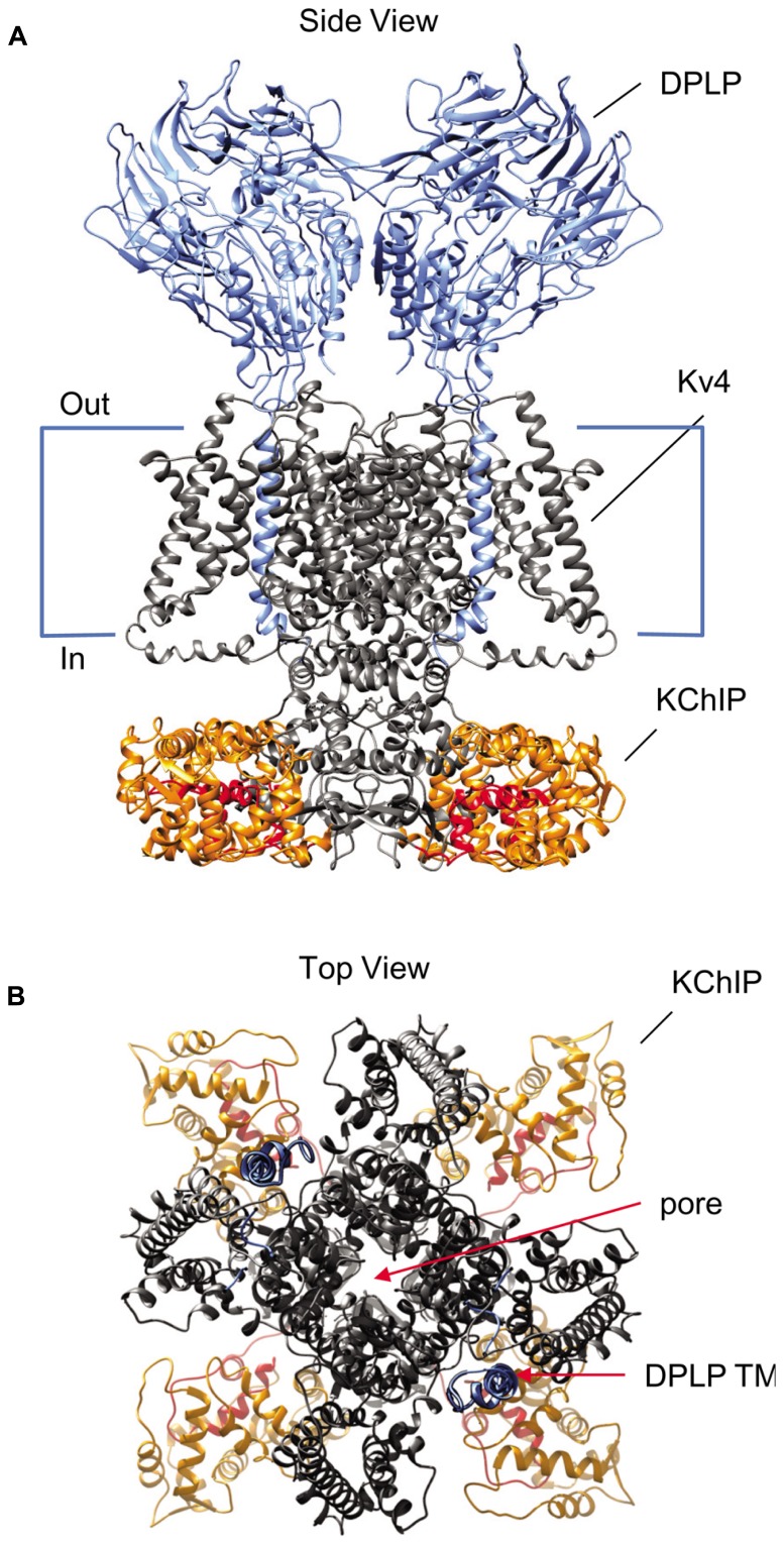
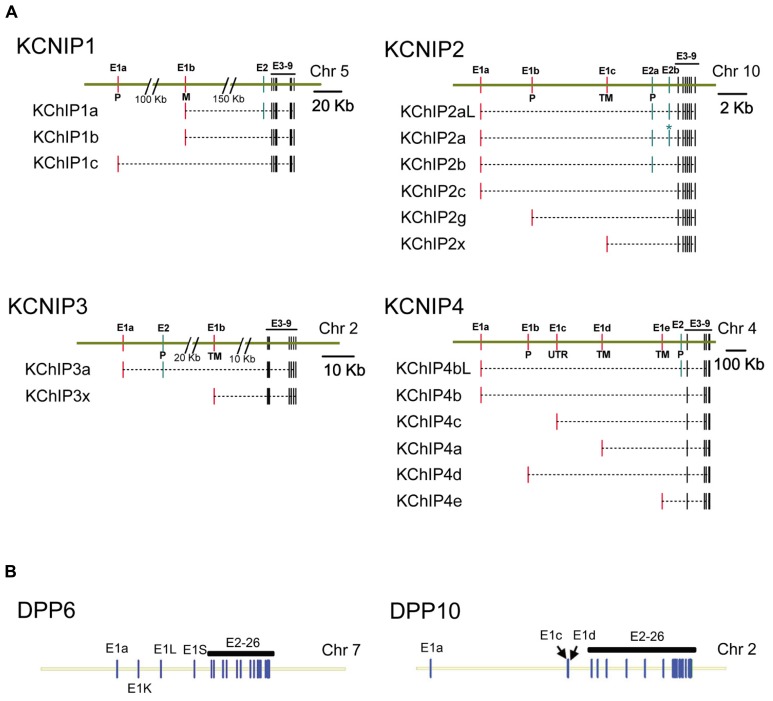
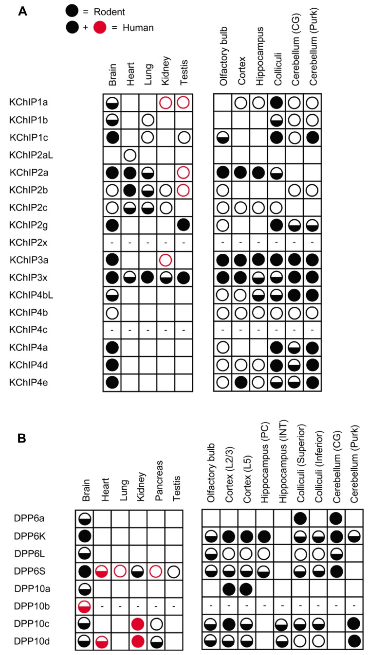
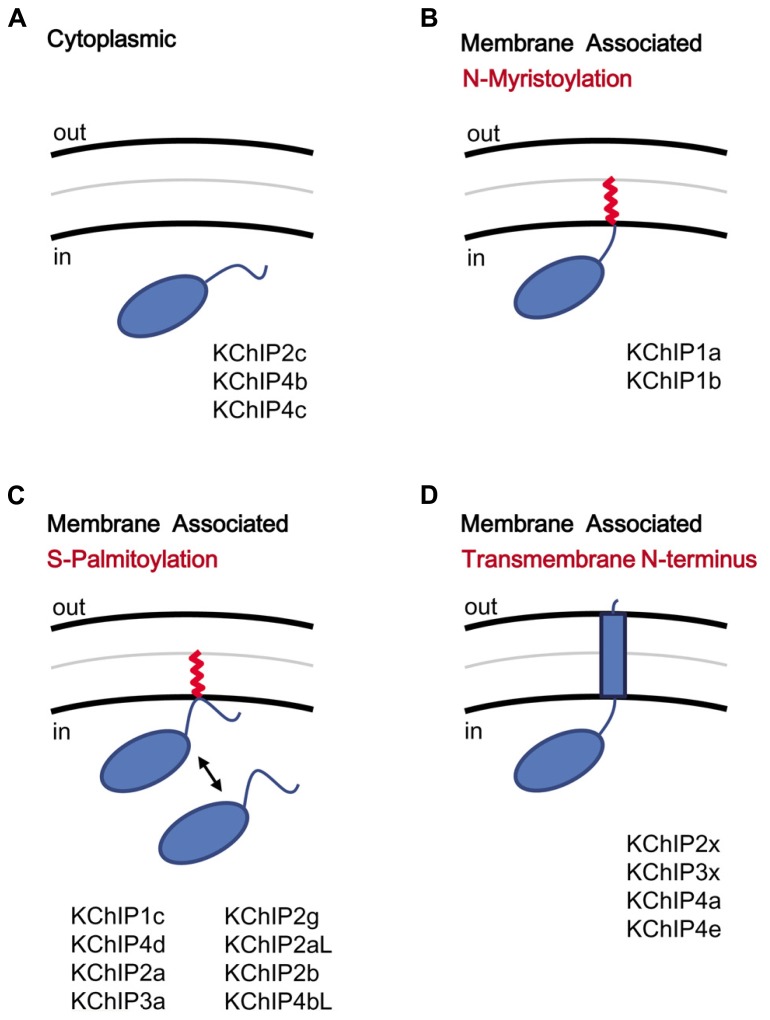
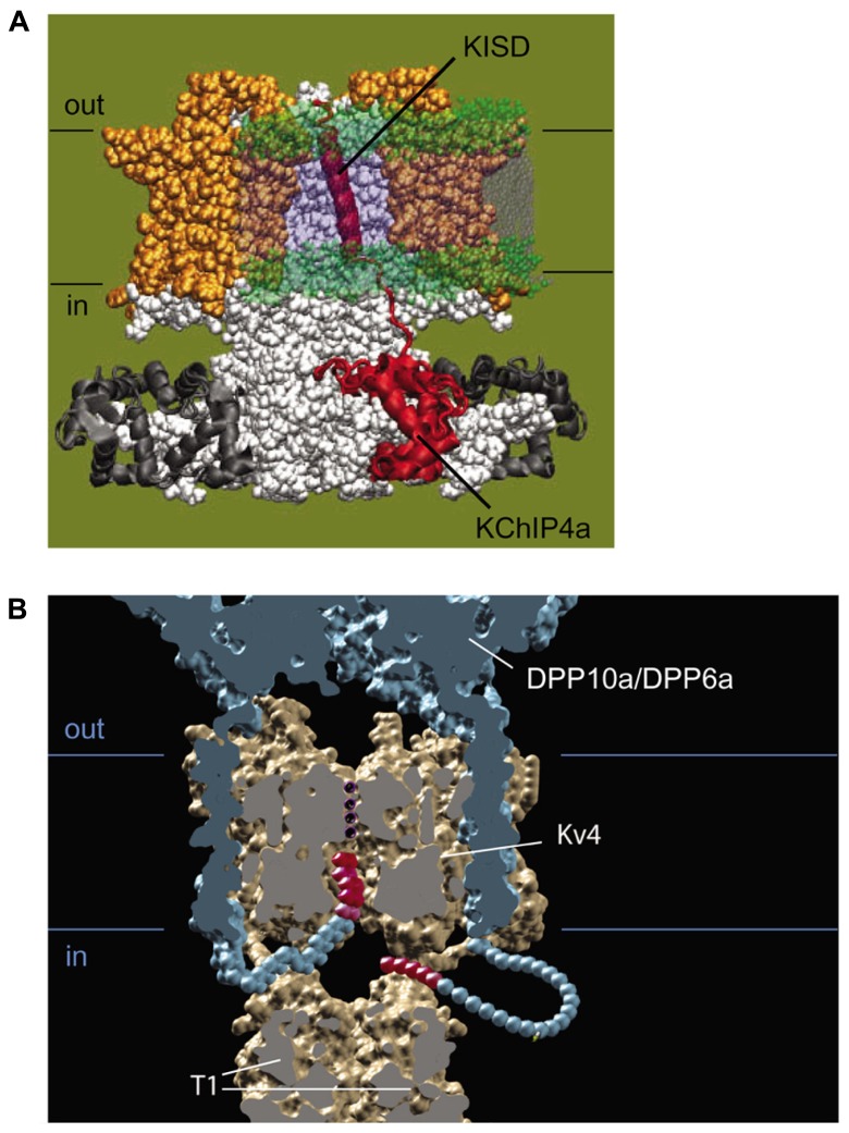
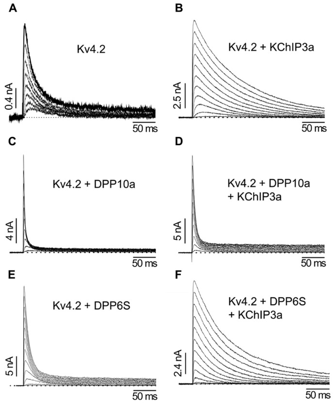
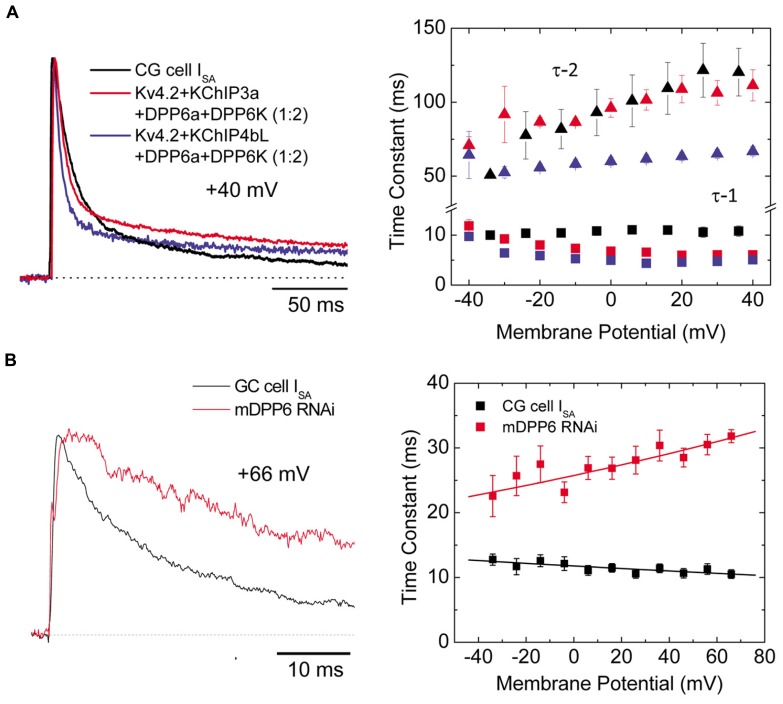
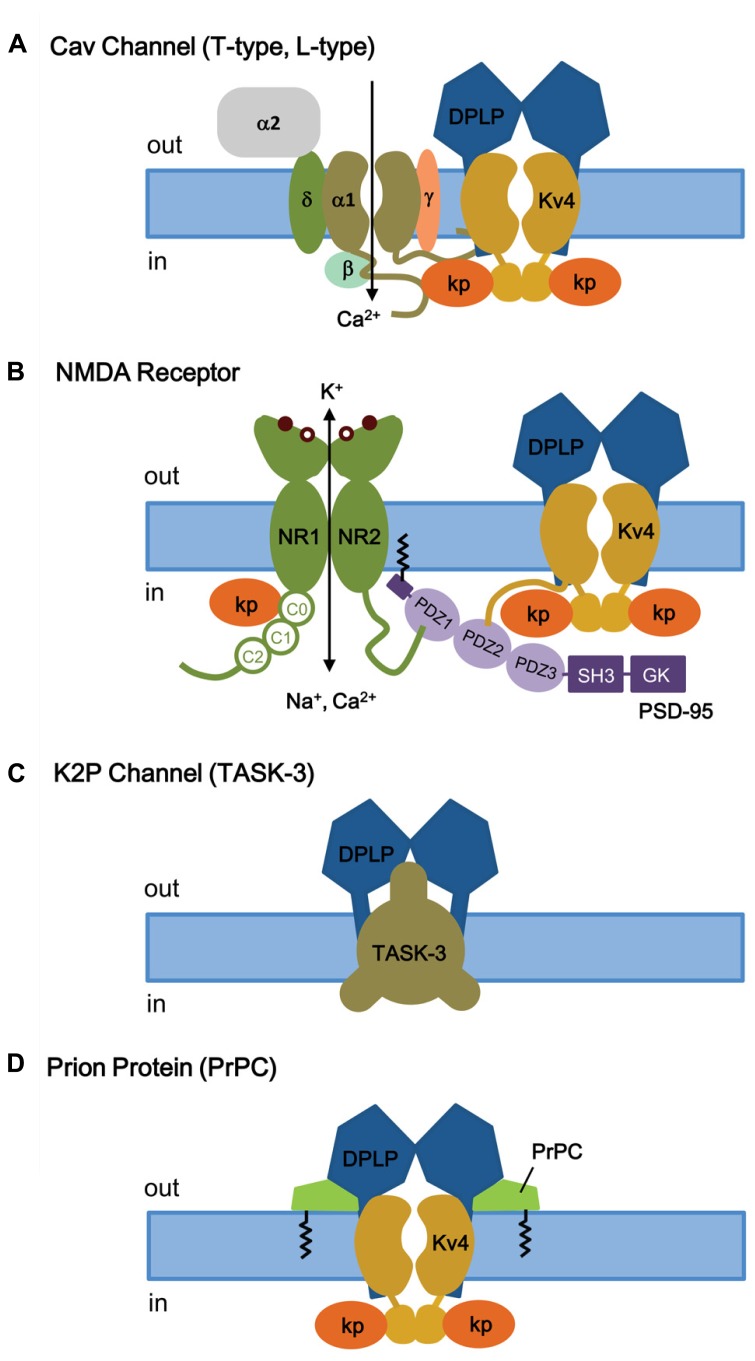
Auxiliary subunits are non-conducting, modulatory components of the multi-protein ion channel complexes that underlie normal neuronal signaling. They interact with the pore-forming α-subunits to modulate surface distribution, ion conductance, and channel gating properties. For the somatodendritic subthreshold A-type potassium (ISA) channel based on Kv4 α-subunits, two types of auxiliary subunits have been extensively studied: Kv channel-interacting proteins (KChIPs) and dipeptidyl peptidase-like proteins (DPLPs). KChIPs are cytoplasmic calcium-binding proteins that interact with intracellular portions of the Kv4 subunits, whereas DPLPs are type II transmembrane proteins that associate with the Kv4 channel core. Both KChIPs and DPLPs genes contain multiple start sites that are used by various neuronal populations to drive the differential expression of functionally distinct N-terminal variants. In turn, these N-terminal variants generate tremendous functional diversity across the nervous system. Here, we focus our review on (1) the molecular mechanism underlying the unique properties of different N-terminal variants, (2) the shaping of native ISA properties by the concerted actions of KChIPs and DPLP variants, and (3) the surprising ways that KChIPs and DPLPs coordinate the activity of multiple channels to fine-tune neuronal excitability. Unlocking the unique contributions of different auxiliary subunit N-terminal variants may provide an important opportunity to develop novel targeted therapeutics to treat numerous neurological disorders.

Keywords: Kv channel-interacting protein; N-terminal variant; auxiliary subunit; dipeptidyl peptidase-like protein; excitability; modulatory mechanism; potassium channel; somatodendritic A-type current.


Figures

FIGURE 1
FIGURE 1
Structural model of the Kv4-KChIP-DPLP supermolecular complex. The structure of the KChIP1-Kv4.3 T1 complex from Pioletti et al. (2006) was docked with the Kv1.2 structure from Long et al. (2007). The structure of DPP6 dimer from Strop et al. (2004) is positioned so that the transmembrane segments are near the voltage-sensing domains of the channel core domains. The structure depicts only two DPP6 molecules, while the experimental evidence suggests that complete complex contains four DPLP subunits (Soh and Goldstein, 2008). (A) Side view of the multi-protein channel complex. Kv4 pore-forming subunit is shown in gray, with the exception of the pre-T1 N-terminal domain which is shown in red. The KChIP molecules are shown in orange; the DPLP molecules, blue. This view clearly shows that KChIP binding sequesters the Kv4 N-terminus. (B) Top view of the complex as seen from the extracellular side. The central pore generated by the Kv4 subunits is indicated and can be easily observed. Note the KChIP molecules (orange) and the DPLP transmembrane domains (TM, blue) reside between the Kv4 voltage sensors with free access to the T1 side windows.
FIGURE 2
FIGURE 2
Genomic structure of KChIP and DPLP genes. Gene structures for Kv4.2 auxiliary subunit proteins show a common core with variable N-terminal exons. (A) KChIP genes show a common set of 3′ exons 3–9 with 1 to 2 alternative 5′ exons used to construct the final mRNA transcript. (B) DPLP genes show a common set of 3′ exons 2–26 with alternative 5′ first exons. Maps were constructed using exon locations in human chromosomes as given in Concensus CDS data base (http://www.ncbi.nlm.nih.gov/CCDS) or determined from published mRNA sequences using Splign (http://www.ncbi.nlm.nih.gov/sutils/splign). Additional splice variants of these genes have been proposed that are not shown. (A) KChIP1: KChIP1a-CCDS34286, KChIP1b-CCDS4374, KChIP1c-CCDS34285. KChIP2: KChIP2al-CCDS7521, KChIP2a-CCDS7522, KChIP2b-CCDS41562, KChIP2c-CCDS7524, KChIP2g-CCDS7525, KChIP2x- Jerng and Pfaffinger (2008). KChIP3: KChIP3a-CCDS2013, KChIP3x-CCDS33245. KChIP4: KChIP4bL-CCDS43216, KChIP4b-CCDS43215, KChIP4c-CCDS47035, KChIP4a-CCDS3428, KChIP4d-CCDS43217, KChIP4e- Jerng and Pfaffinger (2008). (B) DPP6: splicing based on Jerng et al. (2009). DPP10: DPP10a-CCDS46400, DPP10c-CCDS54388, DPP10d-CCDS33278. P, S-palmitoylation; M, N-myristoylation; TM, transmembrane.
FIGURE 3
FIGURE 3
Differential expression of auxiliary subunit N-terminal variants in various tissues and brain regions in mouse and human. (A) Summary of the expression patterns of KChIP N-terminal variants in mouse (black) and human (black and red): KChIP1 (Van Hoorick et al., 2003; Pruunsild and Timmusk, 2005), KChIP2 (Takimoto et al., 2002; Pruunsild and Timmusk, 2005), KChIP3 (Pruunsild and Timmusk, 2005), and KChIP4 (Holmqvist et al., 2002; Pruunsild and Timmusk, 2005). (B) Summary of the expression patterns of DPLP N-terminal variants in mouse (black) and human (black and red): DPP6 (Wada et al., 1992; de Lecea et al., 1994; Kin et al., 2001; Radicke et al., 2005; Nadal et al., 2006; Maffie et al., 2009; Jerng and Pfaffinger, 2012) and DPP10 (Chen et al., 2006a; Takimoto et al., 2006; Jerng et al., 2007). No symbols, no expression; open circle, low expression; half-filled circle, moderate expression; filled circle, high expression; dash, not determined; CG, cerebellar granule cells; Purk, cerebellar Purkinje cells; PC, pyramidal neuron; INT, interneuron; L2/3, layer 2 and 3; L5, layer 5.
FIGURE 4
FIGURE 4
KChIP N-terminal variants and the motifs coding for membrane association. KChIPs can be freely cytoplasmic or membrane associated. (A) Cytoplasmic KChIPs lack the sequence motif to allowing anchoring of the KChIP molecule to the membrane. (B) Membrane association by N-myristoylation. The myristoylate group (red) is attached to a glycine residue at position 2. (C) Membrane association by S-palmitoylation. The palmitoylate group (red) is attached to a reactive cysteine within the N-terminal domain via a reversible reaction and induces membrane association. (D) Membrane association by insertion of a transmembrane N-terminal segment. Multiple N-terminal variants possess a highly hydrophobic segment that traverses the membrane. Aside from functioning as a membrane anchor, the transmembrane segment also modulates surface trafficking and channel gating.
FIGURE 5
FIGURE 5
Molecular mechanism of functional regulation by KChIP4a, DPP6a, and DPP10a. (A) Working molecular model of the transmembrane Kv-channel inactivation suppressor domain (KISD) inserting through the membrane and making contacts with the Kv4 channel core, as adapted from Jerng and Pfaffinger (2008). As reported by Tang et al. (2013), separate domains within the KISD determine ER retention and regulation of channel gating. KChIP4a is shown in red, voltage sensors in yellow, and the remaining Kv4 channels in white. (B) Side view of a model illustrating the basis for fast inactivation mediated by DPP6a and DPP10a. Two of the four subunits have been removed to reveal the inner channel structure, including the permeation pathway with K+, DPLP extracellular and transmembrane domain (blue), and the DPLP N-termini with inactivation particles (red).
FIGURE 6
FIGURE 6
The differential functional effects of DPLP N-terminal variants (Adapted from Jerng et al., 2005). (A) Kv4.2 outward currents elicited by membrane depolarization in CHO cells. (B) After co-expression with KChIP3a, Kv4.2 channels significantly increase in surface expression and exhibit slower inactivation. (C) Similar to KChIP3a, co-expression of Kv4.2 with DPP10a results in a dramatic increase in peak current; however, the inactivation kinetics is markedly accelerated. (D) Channel complexes co-expressing KChIP3a and DPP10a results in currents that are more like that of Kv4.2+DPP10a channels, indicating the dominance of DPP10a-mediated fast inactivation. (E) DPP6-S accelerates inactivation of Kv4.2 channels. (F) Co-expression of DPP6-S with Kv4.2+KChIP3a channels does not produce dramatic acceleration of inactivation observed with DPP10a.
FIGURE 7
FIGURE 7
Ternary channel complex consisting of Kv4.2, KChIP3a, DPP6a, and DPP6K express A-type current similar to ISA from CG cells (Adapted from Nadin and Pfaffinger, 2010; Jerng and Pfaffinger, 2012). (A) Comparison between native ISA from CG cells and reconstituted currents expressed by Kv4.2, KChIP, and DPP6 subunits. Based on real-time RT-PCR results, the ratio between DPP6a and DPP6K was determined to be 1-to-2 (Jerng and Pfaffinger, 2012). Native and reconstituted current traces elicited by +40 mV depolarizations are quite similar (left panel). The incorporation of KChIP3a verses KChIP4bL produces negligible effect on overall current waveforms. The overall inactivation is best described by the sum of two exponential components, and the two corresponding time constants (τ -1, τ -2) have different dependence on membrane potential (right panel). (B) In the absence of DPP6, ISA inactivation significantly slows (left panel). The loss of DPP6 altered the inactivation-voltage profile (right panel). Control ISA shows inactivation with time constant relatively insensitive to depolarization. After DPP6 knockdown by RNAi, inactivation time constant slows with increasing depolarization. Note that panels A and B have separate legends.
FIGURE 8
FIGURE 8
KChIP and DPLP are also involved in binding and regulating other channels and receptors. (A) T- and L-type Cav channels. Depolarization-activated Cav channels, like ISA channels, are multi-protein complexes. The cytoplasmic N- and C-termini of Cav channels have been shown to interact with the ISA channel complex, via Kv4 or KChIP subunits. (B) NMDA receptor. The NMDA receptors are opened by 2 simultaneous events: activation by extracellular ligand binding and depolarization-mediated relief of block by Mg2+ (coincidence detector). Both NMDA receptor and ISA channels are localized post-synoptically, and KChIPs have been shown to regulate NMDA receptor activities. (C) TASK-3 two-pore K+ channels. DPP6 co-assembles with TASK-3 channels in the dendritic membrane to regulate membrane excitability. (D) Prion proteins (PrPC) bind to DPP6, thereby regulating ISA expression and gating. Kp, KChIP; NR1, glycine-binding subunit; NR2, glutamate-binding subunit; SH3, SRC homology 3 domain; GK, guanylate kinase.

Similar articles

Cited by

References

    1. Aimond F., Kwak S. P., Rhodes K. J., Nerbonne J. M. (2005). Accessory Kvbeta1 subunits differentially modulate the functional expression of voltage-gated K+ channels in mouse ventricular myocytes. Circ. Res. 96 451–458 10.1161/01.RES.0000156890.25876.63 - DOI - PubMed
    1. Alexander J. C., McDermott C. M., Tunur T., Rands V., Stelly C., Karhson D., et al. (2009). The role of calsenilin/DREAM/KChIP3 in contextual fear conditioning. Learn. Mem. 16 167–177 10.1101/lm.1261709 - DOI - PMC - PubMed
    1. Allen M., Heinzmann A., Noguchi E., Abecasis G., Broxholme J., Ponting C. P., et al. (2003). Positional cloning of a novel gene influencing asthma from chromosome 2q14. Nat. Genet. 35 258–263 10.1038/ng1256 - DOI - PubMed
    1. Aller M. I., Veale E. L., Linden A. M., Sandu C., Schwaninger M., Evans L. J., et al. (2005). Modifying the subunit composition of TASK channels alters the modulation of a leak conductance in cerebellar granule neurons. J. Neurosci. 25 11455–11467 10.1523/JNEUROSCI.3153-05.2005 - DOI - PMC - PubMed
    1. Aller M. I., Wisden W. (2008). Changes in expression of some two-pore domain potassium channel genes (KCNK) in selected brain regions of developing mice. Neuroscience 151 1154–1172 10.1016/j.neuroscience.2007.12.011 - DOI - PubMed

LinkOut - more resources