. 2014 Mar 26:8:177.
doi: 10.3389/fnhum.2014.00177. eCollection 2014.

Enhanced peripheral visual processing in congenitally deaf humans is supported by multiple brain regions, including primary auditory cortex

Affiliations

Enhanced peripheral visual processing in congenitally deaf humans is supported by multiple brain regions, including primary auditory cortex

Gregory D Scott et al. Front Hum Neurosci. .

Abstract

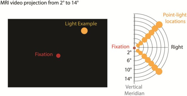
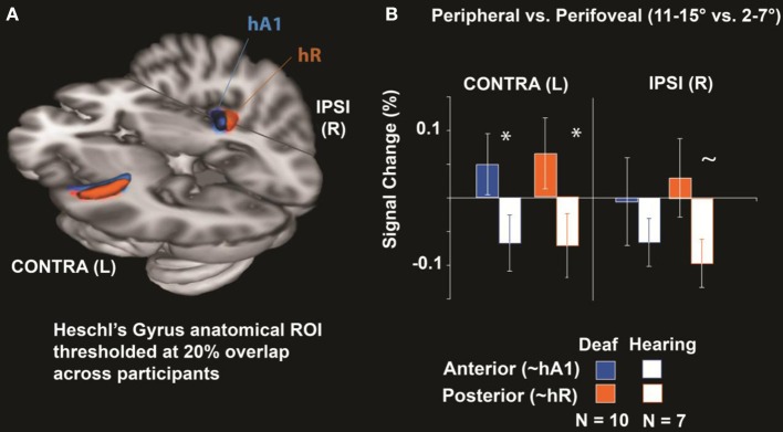
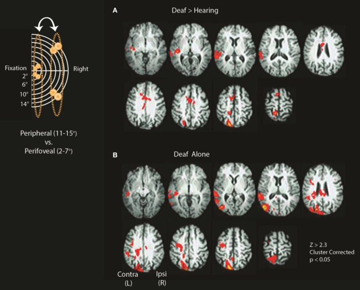
Brain reorganization associated with altered sensory experience clarifies the critical role of neuroplasticity in development. An example is enhanced peripheral visual processing associated with congenital deafness, but the neural systems supporting this have not been fully characterized. A gap in our understanding of deafness-enhanced peripheral vision is the contribution of primary auditory cortex. Previous studies of auditory cortex that use anatomical normalization across participants were limited by inter-subject variability of Heschl's gyrus. In addition to reorganized auditory cortex (cross-modal plasticity), a second gap in our understanding is the contribution of altered modality-specific cortices (visual intramodal plasticity in this case), as well as supramodal and multisensory cortices, especially when target detection is required across contrasts. Here we address these gaps by comparing fMRI signal change for peripheral vs. perifoveal visual stimulation (11-15° vs. 2-7°) in congenitally deaf and hearing participants in a blocked experimental design with two analytical approaches: a Heschl's gyrus region of interest analysis and a whole brain analysis. Our results using individually-defined primary auditory cortex (Heschl's gyrus) indicate that fMRI signal change for more peripheral stimuli was greater than perifoveal in deaf but not in hearing participants. Whole-brain analyses revealed differences between deaf and hearing participants for peripheral vs. perifoveal visual processing in extrastriate visual cortex including primary auditory cortex, MT+/V5, superior-temporal auditory, and multisensory and/or supramodal regions, such as posterior parietal cortex (PPC), frontal eye fields, anterior cingulate, and supplementary eye fields. Overall, these data demonstrate the contribution of neuroplasticity in multiple systems including primary auditory cortex, supramodal, and multisensory regions, to altered visual processing in congenitally deaf adults.

Keywords: Heschl's gyrus; auditory cortex; deaf; fMRI; human; visual attention.


Figures

Figure 1
Figure 1
Stimulus presentation and Apparatus. We used a standard MRI video projection system, back projected to a screen viewed by the participants through a monitor. A red fixation point was displayed continuously at center and single point yellow light stimuli were presented in the upper or lower right visual field at polar eccentricities ranging from 2 to 15°. Standard stimuli (85%) were flashed at 8 Hz and target stimuli requiring a button press (15%) were flashed at 14 Hz.
Figure 2
Figure 2
Heschl's Gyrus Region of interest analysis. (A) Anatomical Heschl's gyrus ROIs drawn on individual structural brain images divided along the first principal component axis into an anterior and posterior subregion to approximate primate primary-auditory areas A1 and R respectively (as defined tonotopically in hearing adults; Da Costa et al., 2011), shown here thresholded at 20% overlap across participants. (B) Differences between peripheral (11–15°) and perifoveal locations (2–7°) were significantly larger in deaf than hearing participants for anterior and posterior Heschl's gyrus contralateral to visual stimulation (*p < 0.05) and tended to be larger in deaf participants posterior Heschl's gyrus ipsilateral to stimulation (~ p < 0.10). Error bars represent ± s.e.m.
Figure 3
Figure 3
Differences between deaf and hearing for peripheral vs. perifoveal stimulation. Inset shows a schematic location of stimuli included in the contrasts (11–15° vs. 2–7°). (A) Deaf > Hearing and (B) Deaf alone. See Tables 1, 2 for a summary of significant clusters and corresponding atlas-based descriptions.

References

    1. Allman B., Keniston L., Meredith M. (2009). Adult deafness induces somatosensory conversion of ferret auditory cortex. Proc. Natl. Acad. Sci. U.S.A. 106, 5925–5930 10.1073/pnas.0809483106 - DOI - PMC - PubMed
    1. Auer E. T., Jr., Bernstein L., Sungkarat W., Singh M. (2007). Vibrotactile activation of the auditory cortices in deaf versus hearing adults. Neuroreport 18, 645–648 10.1097/WNR.0b013e3280d943b9 - DOI - PMC - PubMed
    1. Bavelier D., Brozinsky C., Tomann A., Mitchell T., Neville H., Liu G. (2001). Impact of early deafness and early exposure to sign language on the cerebral organization for motion processing. J. Neurosci. 21, 8931–8942 - PMC - PubMed
    1. Bavelier D., Corina D., Jezzard P., Clark V., Karni A., Lalwani A., et al. (1998). Hemispheric specialization for English and ASL: Left invariance - right variability. Neuroreport 9, 1537–1542 10.1097/00001756-199805110-00054 - DOI - PubMed
    1. Bavelier D., Dye M., Hauser P. (2006). Do deaf individuals see better? Trends Cogn. Sci. 10, 512–518 10.1016/j.tics.2006.09.006 - DOI - PMC - PubMed

LinkOut - more resources