Review
. 2014 Mar 26:5:123.
doi: 10.3389/fimmu.2014.00123. eCollection 2014.

Structural basis for recognition of cellular and viral ligands by NK cell receptors

Affiliations
Review

Structural basis for recognition of cellular and viral ligands by NK cell receptors

Yili Li et al. Front Immunol. .

Abstract

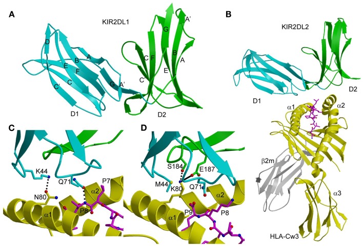
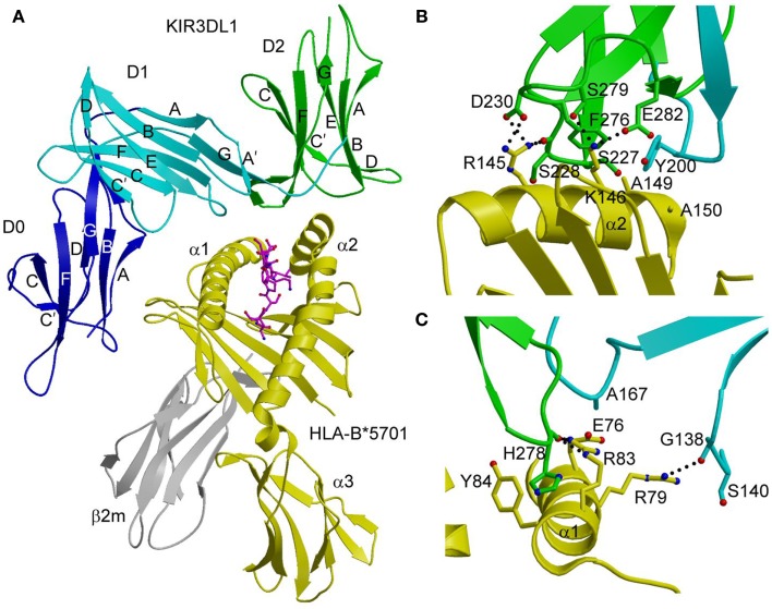
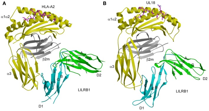
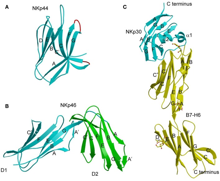
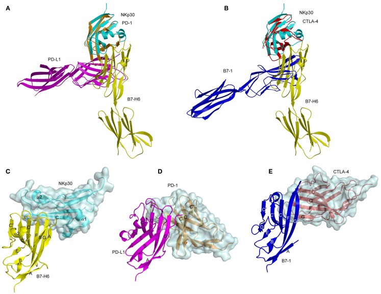
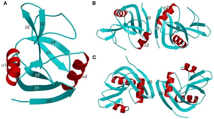
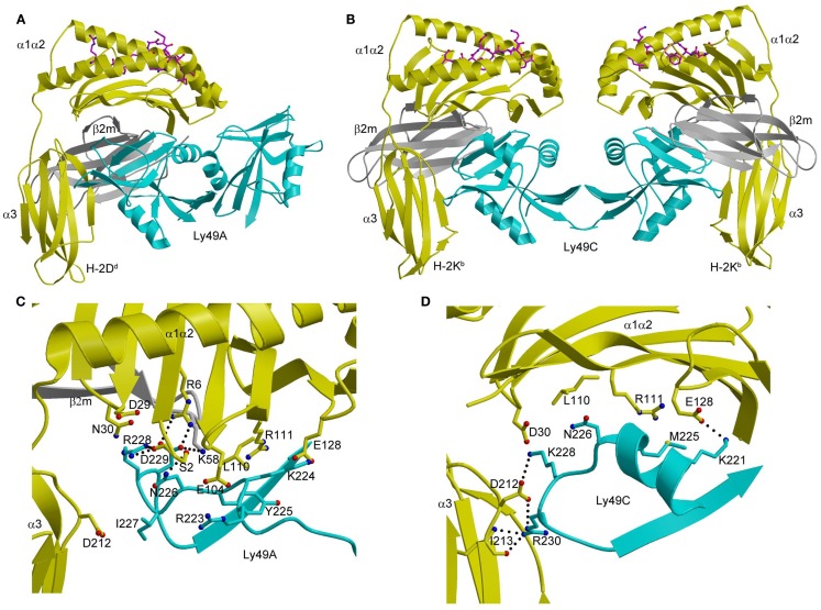
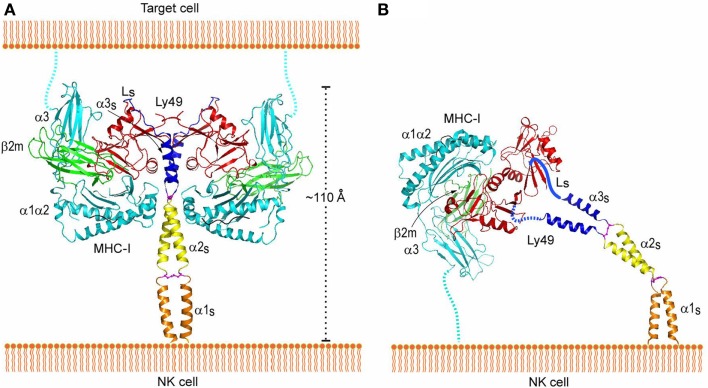
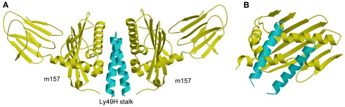
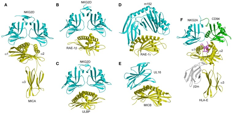
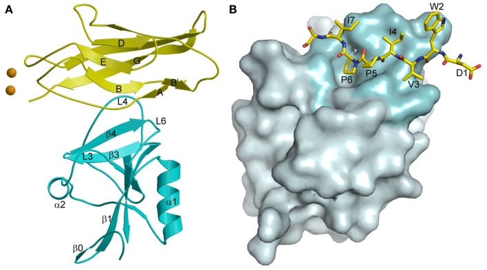
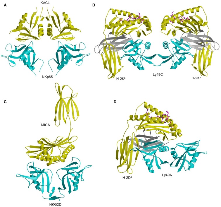
Natural killer (NK) cells are key components of innate immune responses to tumors and viral infections. NK cell function is regulated by NK cell receptors that recognize both cellular and viral ligands, including major histocompatibility complex (MHC), MHC-like, and non-MHC molecules. These receptors include Ly49s, killer immunoglobulin-like receptors, leukocyte immunoglobulin-like receptors, and NKG2A/CD94, which bind MHC class I (MHC-I) molecules, and NKG2D, which binds MHC-I paralogs such as the stress-induced proteins MICA and ULBP. In addition, certain viruses have evolved MHC-like immunoevasins, such as UL18 and m157 from cytomegalovirus, that act as decoy ligands for NK receptors. A growing number of NK receptor-ligand interaction pairs involving non-MHC molecules have also been identified, including NKp30-B7-H6, killer cell lectin-like receptor G1-cadherin, and NKp80-AICL. Here, we describe crystal structures determined to date of NK cell receptors bound to MHC, MHC-related, and non-MHC ligands. Collectively, these structures reveal the diverse solutions that NK receptors have developed to recognize these molecules, thereby enabling the regulation of NK cytolytic activity by both host and viral ligands.

Keywords: KIR; Ly49; MHC; NK receptor; NKG2; structure; virus.


Figures

Figure 1
Figure 1
Three-dimensional structures of KIR2DL and KIR2DL–HLA-C complexes. (A) Ribbon diagram of KIR2DL1 (PDB accession code 1NKR). The D1 domain is cyan; D2 is green. The secondary structural elements are labeled. (B) Ribbons diagram of KIR2DL2 bound to HLA-Cw3 (1EFX). The α1, α2, and α3 domains of the HLA-Cw3 heavy chain are yellow; β2m is gray; the peptide is magenta. (C) Basis for allelic specificity and peptide selectivity of KIR2D receptors. The dotted lines represent hydrogen bonds formed by Asn80 of HLA-Cw3 with Lys44 of KIR2DL2, and by Gln71 of HLA-Cw3 with P8 of the peptide. (D) Interactions of Lys80 of HLA-Cw4 (yellow) with specificity-determining residues of KIR2DL1 (D1 domain in cyan, D2 domain in green) in the KIR2DL1–HLA-Cw4 complex (1IM9). The solid line represents a salt bridge.
Figure 2
Figure 2
Structure of the KIR3DL1–HLA-B*5701 complex. (A) Ribbon diagram of KIR3DL1 bound to HLA-B*5701 (3VH8). The orientation of the MHC-I ligand is similar to that of HLA-Cw3 in the KIR2DL2–HLA-Cw3 complex (Figure 1B). The HLA-B*5701 heavy chain is yellow; β2m is gray; the peptide is magenta. The KIR3DL1 D0 domain is dark blue; D1 is cyan; D2 is green. The secondary structural elements of KIR3DL1 are labeled. (B) Contacts between KIR3DL1 and the HLA-B*5701 α2 helix. The D2 domain mainly interacts with HLA-B*5701 residues 142–151, which display limited polymorphism among HLA-B alleles. At the center of the D2–HLA-B*5701 interface, KIR3DL1 residues Tyr200 and Phe276 form an aromatic cluster that converges on the α2 helix. (C) Contacts between KIR3DL1 and the HLA-B*5701 α1 helix. KIR3DL1 recognizes HLA allotypes that contain the Bw4 epitope-defining residues 77–83 on the α1 helix, which likely accounts for the allelic specificity of KIR3DLs.
Figure 3
Figure 3
Interaction of LILRB1 with MHC-I and a viral MHC-I mimic. (A) Structure of LILRB1 bound to HLA-A2 (1P7Q). The α1, α2, and α3 domains of the HLA-A2 heavy chain are yellow; β2m is gray; the peptide is magenta. The D1 and D2 domains of LILRB1 are colored in cyan and green, respectively. The secondary structural elements of LILRB1 are labeled. (B) Structure of LILRB1 bound to the HCMV MHC-I mimic UL18 (3D2U).
Figure 4
Figure 4
Natural cytotoxicity receptors. (A) Structure of NKp44 (1HKF). The β-strands are labeled. The CC′ and FG loops, drawn in red, define a positively charged surface groove that may serve as a binding site for anionic ligands. (B) Structure of NKp46 (1P6F). D1 is cyan; D2 is green. (C) Structure of NKp30 bound to its tumor cell ligand B7-H6 (3PV6). N-linked glycans at B7-H6 residues Asn43 and Asn57 in the V-like domain and Asn208 in the C-like domain are shown in ball-and-stick representation.
Figure 5
Figure 5
Comparison of the NKp30–B7-H6, PD-1–PD-L1, and CTLA-4–B7-1 complexes. (A) Overlay of the NKp30–B7-H6 (3PV6) and PD-1–PD-L1 (3BIK) complexes by superposing NKp30 (cyan) onto PD-1 (brown). B7-H6 is yellow; PD-L1 is violet. (B) Overlay of the NKp30–B7-H6 and CTLA-4–B7-1 (1I8L) complexes by superposing NKp30 (cyan) onto CTLA-4 (red). B7-H6 is yellow; B7-1 is dark blue. (C–E) Docking modes in the NKp30–B7-H6, PD-1–PD-L1, and CTLA-4–B7-1 complexes. The three complexes were overlaid by superposing the IgV domains of B7-H6 (yellow), PD-1-L1 (violet), and B7-1 (dark blue), then translated horizontally for viewing.
Figure 6
Figure 6
Structures of Ly49 NK receptors. (A) Ribbon drawing of the Ly49A C-type lectin-like domain (1QO3). Secondary structure elements are labeled. β-strands and loops are cyan; α-helices are red. (B) Structure of the “closed” Ly49A homodimer. Secondary structure elements that participate in formation of the dimer interface are labeled. The α2 helices are juxtaposed. (C) Structure of the “open” Ly49C homodimer (3C8J). The α2 helices do not make contact across the dimer interface.
Figure 7
Figure 7
Structures of Ly49–MHC-I complexes. (A) Ribbon diagram of Ly49A bound to H-2Dd (1QO3). The α1, α2, and α3 domains of the MHC-I heavy chain are yellow; β2m is gray; the peptide is magenta; the Ly49A dimer is cyan. In this view, the complex is oriented with the H-2Dd molecule on the target cell at the bottom; the Ly49A homodimer reaches H-2Dd from an opposing NK cell at the top, to which it is connected via stalk regions projecting down to the N-termini of the subunits. (B) Structure of Ly49C in complex with H-2Kb (3C8K). (C) The Ly49A–H-2Dd interface, highlighting interactions made by residues 211–231 of Ly49A. (D) The Ly49C–H-2Kb interface, showing interactions made by the corresponding region of Ly49C. The side chains of interacting residues are drawn in ball-and-stick representation, with carbon atoms in cyan (Ly49A or Ly49C), yellow (H-2Dd or H-2Kb), or gray (β2m), oxygen atoms in red, and nitrogen atoms in blue. Hydrogen bonds are represented by dotted lines.
Figure 8
Figure 8
Interaction of Ly49 receptors with MHC-I in trans and cis. (A) Trans interaction of an Ly49 receptor with two MHC-I molecules, based on the structures of Ly49L (3G8L) and the Ly49C–H-2Kb complex (3C8K). The α1, α2, and α3 domains of the MHC-I heavy chain are cyan; β2m is green; Ly49 NKD is red; helix α3S of the Ly49 stalk and loop LS connecting α3S to the NKD are blue; the disulfide bond linking the α3S helices is magenta. The predicted α1S and α2S helices of the stalk are orange and yellow, respectively, with the disulfide bond in magenta. The Ly49 homodimer on the NK cell binds two MHC-I molecules on the target cell. To bind in trans, the stalks must adopt a backfolded conformation, as the N-termini of the Ly49 monomers point away from the NK cell membrane (Ly49s are type II transmembrane proteins). (B) Cis interaction of Ly49 with MHC-I, based on the structure of Ly49L and the Ly49A–H-2Dd complex (1QO3). The LS loops connecting the α3S helices to the NKDs are drawn arbitrarily. The Ly49 homodimer binds one MHC-I molecule on the NK cell itself. In this case, the stalks must assume an extended conformation, as the N-termini of the Ly49 monomers point toward the NK cell. Reproduced with permission from Immunity (106).
Figure 9
Figure 9
Structure of m157 bound to the stalk region of Ly49H. (A) Side view of the Ly49H–m157 complex (4JO8), in which two m157 monomers (yellow) engage the Ly49H stalk (cyan). Only part of the helical stalk region of Ly49H (the α3s segment) was visible in electron density. The rest of the Ly49H stalk (segments α1s and α2s) and the NKD were not resolved in the structure. (B) Top view of the Ly49H–m157 complex, in which the helical stalks of Ly49H lie across the α1/α2 platform of m157.
Figure 10
Figure 10
Structures of NKG2D and NKG2A/CD94 complexes. (A) The human NKG2D–MICA complex (1HYR). The two subunits of the NKG2D homodimer are cyan; MICA is yellow. (B) The mouse NKG2D–RAE-1β complex (1JSK). (C) The human NKG2D–ULBP3 complex (1KCG). (D) Structure of the MCMV immunoevasin m152 bound to the NKG2D ligand RAE-1γ (4G59). (E) Structure of the Ig-like HCMV immunoevasin UL16 bound to MICA (2WY3). (F) The human NKG2A/CD94–HLA-E complex (3CDG). The NKG2A and CD94 subunits of the NKG2A/CD94 heterodimer are cyan and green, respectively.
Figure 11
Figure 11
Cadherin recognition by KLRG1. (A) Structure of KLRG1 in complex with the membrane-distal D1 domain of E-cadherin (3FF8). KLRG1 is cyan and E-cadherin is yellow. Secondary structure elements are labeled. Bound Ca2+ ions are drawn as brown spheres. (B) The KLRG1–E-cadherin binding interface. The KLRG1 molecular surface is shown in gray with the region contacting E-cadherin colored cyan. Residues 1–8 of E-cadherin are drawn in stick format and labeled.
Figure 12
Figure 12
Structure of the NKp65–KACL complex and comparison with other NKC-encoded receptor complexes. (A) Structure of the human NKp65–KACL complex (4IOP). NKp65 is cyan and the KACL dimer is yellow. (B) Structure of the Ly49C–H-2Kb complex (3C8K). The Ly49C dimer is cyan, the H-2Kb heavy chain is yellow, and β2m is gray. (C) Structure of the NKG2D–MICA complex (1HYR). The NKG2D dimer is cyan and MICA is yellow. (D) Structure of the Ly49A–H-2Dd complex (1QO3). The Ly49A dimer is cyan, the H-2Dd is heavy chain is yellow, and β2m is gray.

Similar articles

Cited by

References

    1. Di Santo JP. Natural killer cells: diversity in search of a niche. Nat Immunol (2008) 9:473–510.1038/ni.f.201 - DOI - PubMed
    1. Vivier E, Tomasello E, Baratin M, Walzer T, Ugolini S. Functions of natural killer cells. Nat Immunol (2008) 9:503–1010.1038/ni1582 - DOI - PubMed
    1. Caliguiri MA. Human natural killer cells. Blood (2008) 112:461–910.1182/blood-2007-09-077438 - DOI - PMC - PubMed
    1. Bryceson YT, Chiang SC, Darmanin S, Fauriat C, Schlums H, Theorell J, et al. Molecular mechanisms of natural killer cell activation. J Innate Immun (2011) 3:216–2610.1159/000325265 - DOI - PubMed
    1. Vivier E, Raulet DH, Moretta A, Caliguiri MA, Zitvogel L, Lanier LL, et al. Innate or adaptive immunity? The example of natural killer cells. Science (2011) 331:44–910.1126/science.1198687 - DOI - PMC - PubMed