. 2013 Feb 27;5(3):24.

Immune cell infiltration patterns and survival in head and neck squamous cell carcinoma

Affiliations

Immune cell infiltration patterns and survival in head and neck squamous cell carcinoma

Sm Russell et al. Head Neck Oncol. .

Abstract

Purpose: This study examines the tumour-host immune interactions in head and neck squamous cell carcinoma (HNSCC) and their relationship to human papillomavirus (HPV) infectivity and patient survival.

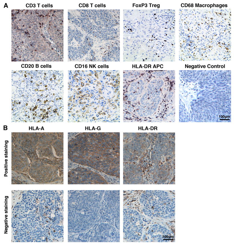
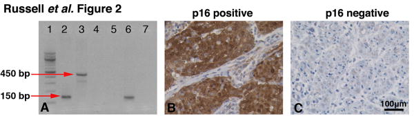
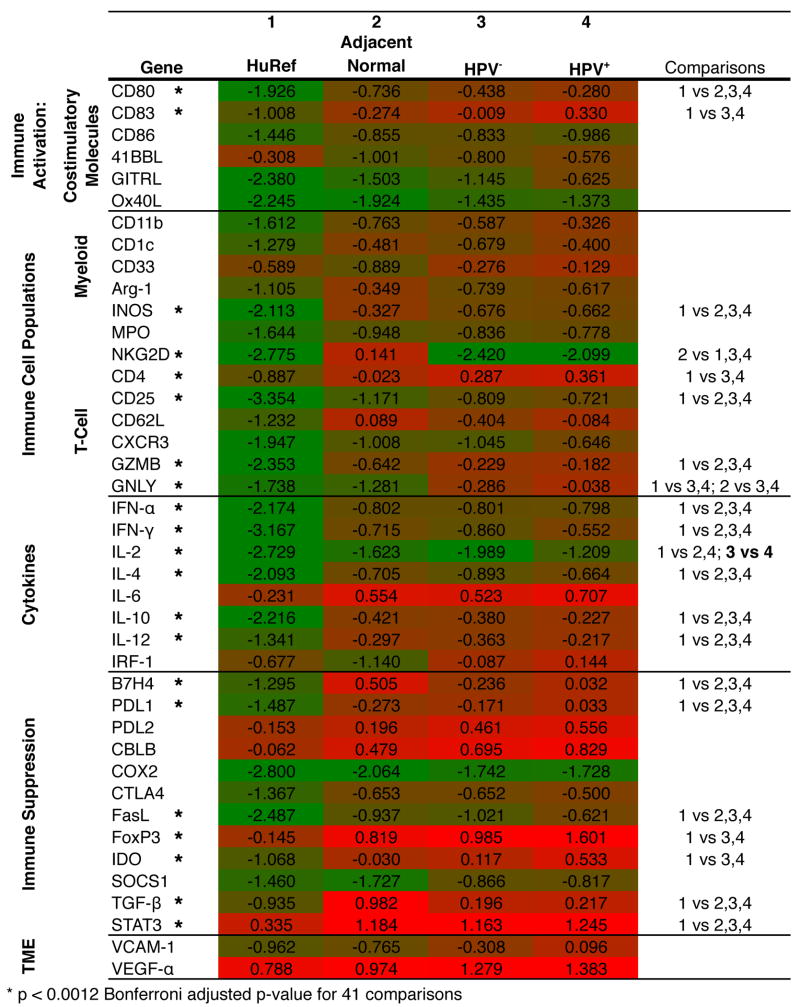
Methods: The adaptive and innate immune profile of surgical tumour specimens obtained from HNSCC patients was determined using qRT-PCR and immunohistochemistry. Intratumoural and invading margin leukocyte populations (CD3, CD8, CD16, CD20, CD68, FoxP3 and HLA-DR) were quantified and compared with patient disease-specific survival. Additionally, the expression of 41 immune activation- and suppression-related genes was evaluated in the tumour microenvironment. Tumour cells were also assessed for expression of HLA-A, HLA-G and HLA-DR. HPV infectivity of tumour biopsies was determined using HPV consensus primers (MY09/MY11 and GP5+/GP6+) and confirmed with p16 immunohistochemistry.

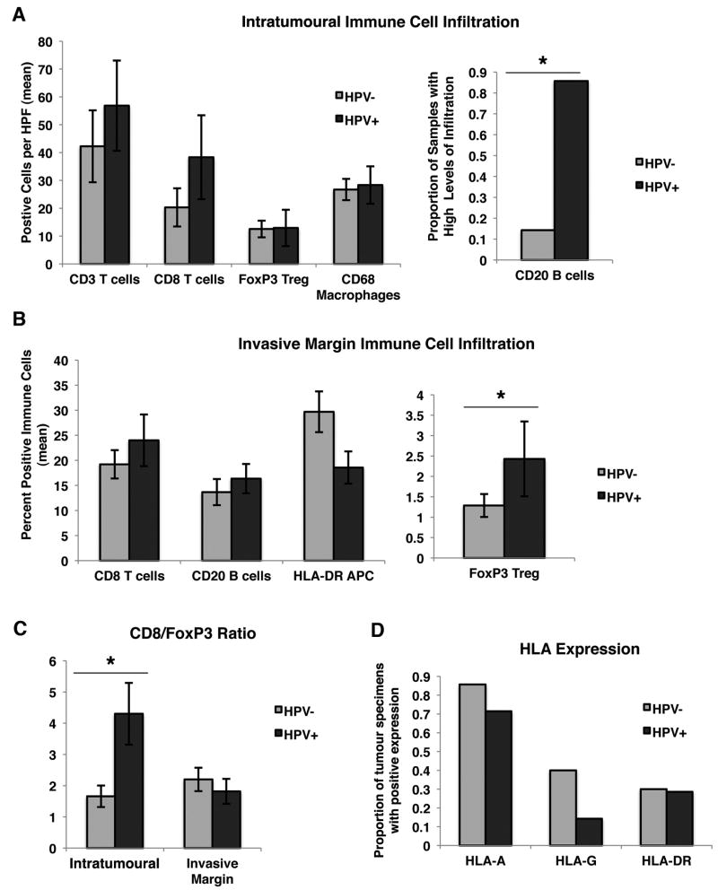
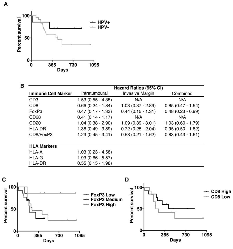
Results: HPV+ patient samples showed a significantly increased infiltration by intratumoural CD20+ B cells, as well as by invasive margin FoxP3+Treg, compared with HPV- patient samples. There was also a trend towards increased intratumoural CD8+ T cells and HLA-G expression on tumour cells in HPV+ samples. qRT-PCR data demonstrated a general pattern of increased immune activation and suppression mechanisms in HPV+ samples. Additionally, a combined score of intratumoural and invasive margin FoxP3 infiltration was significantly associated with disease-specific survival (P < 0.05).

Conclusions: These data demonstrate significant differences in the immune cell profile of HPV+ and HPV- HNSCC. This study identifies several possible targets for immunotherapy and possible prognostic markers (FoxP3 and HLA-G) that may be specific to HNSCC.


Figures

Figure 1
Figure 1
IHC staining of tumour specimens for (a) immune cell populations: CD3+ T cells, CD8+ T cells, FoxP3+Treg (nuclear stain), CD20+ B cells, CD16+ NK cells, CD68+ macrophages and HLA-DR+ APCs on patient tumour specimens (200× original magnification) or (b) MHC class I markers: HLA-A, HLA-G and HLA-DR (top row shows positively-stained tumours, bottom row shows negatively-stained tumours; 200× original magnification).
Figure 2
Figure 2
HPV screening of tumour specimens. Samples were screened for HPV infection using PCR, along with confirmation by p16 staining. (a) Sample PCR results demonstrating amplification of a 150-bp product (GP5+/GP6+) in lane 2 and a 450-bp product (MY09/MY11) in lane 3; HeLa cell line was used as a positive control. The negative control (H2O) for each primer set can be seen in lanes 4 and 5. An HPV+ patient sample loaded in lanes 6 and 7 demonstrates amplification using GP5+/GP6+ primers (Lane 6), but the sample does not show amplification using MY09/MY11 primers (Lane 7). (b) p16 staining on an HPV+ and (c) HPV patient sample (200× original magnification).
Figure 3
Figure 3
Pattern of immune gene expression in HPV+ and HPV HNSCC tumours. A heatmap displaying gene expression data for HuRNA, adjacent normal tissue specimens, HPV patient tumour samples and HPV+ patient tumour samples. Gene-specific amplification values were normalized to GAPDH within each sample, and the mean of the log10% GAPDH for each group is shown (green shading indicates GAPDH = 0.001, red shading indicates % GAPDH = 10).
Figure 4
Figure 4
Effect of HPV infection on tumour infiltration by immune cells. (a) Intratumoural immune cell infiltration in HPV+ and HPV patient tumour samples. The mean number of positive cells per HPF is shown +SEM. CD20 was coded as a categorical variable, and the proportion of samples with high levels of infiltration (greater than median) is shown. (b) Invasive margin immune cell infiltration in HPV+ and HPV patient tumour samples. The mean percentage of positive immune cells is displayed, as well as the SEM. FoxP3 was coded on a categorical scale of 0–3 (by quartile). (c) The ratio of CD8+ T cells to FoxP3+Treg intratumourally and at the invasive margin. (d) The proportion of patient samples positive (>10% of nucleated cells) for HLA-A, HLA-G and HLA-DR. For a–d, * indicates P < 0.05.
Figure 5
Figure 5
Relation of immune infiltrates with HNSCC-specific mortality. (a) Kaplan–Meier curve of survival in HPV+ versus HPV patients. (b) HRs comparing samples with higher levels of immune cell infiltration (top 50%) to those with lower levels of immune cell infiltration (bottom 50%). HRs are reported for intratumoural, invasive margin (peritumoural) and combined populations. HR < 1 indicates decreased and HR > 1 indicates increased HNSCC-specific mortality. (c) Kaplan–Meier curve of survival in subjects with high, moderate or low levels of FoxP3 expression (high = high invasive margin and tumour cell nest infiltration; moderate = high infiltration in one location, low in the other and low = low invasive margin and tumour cell infiltration) (P < 0.05). (d) Kaplan–Meier curve of survival in subjects relative to the extent of intratumoural CD8+ infiltrate.

Similar articles

Cited by

References

    1. Pai SI, Westra WH. Molecular pathology of head and neck cancer: implications for diagnosis, prognosis, and treatment. Annu Rev Pathol. 2009;4:49–70. - PMC - PubMed
    1. Jemal A, Siegel R, Xu J, Ward E. Cancer statistics, 2010. CA Cancer J Clin. 2010 Sep-Oct;60(5):277–300. - PubMed
    1. Goon PK, Stanley MA, Ebmeyer J, Steinsträsser L, Upile T, Jerjes W. HPV & head and neck cancer: a descriptive update. Head Neck Oncol. 2009 Oct;1:36–43. - PMC - PubMed
    1. Dayyani F, Etzel CJ, Liu M, Ho CH, Lippman SM, Tsao AS. Meta-analysis of the impact of human papillomavirus (HPV) on cancer risk and overall survival in head and neck squamous cell carcinomas (HNSCC) Head Neck Oncol. 2010 Jun;2:15. - PMC - PubMed
    1. Duray A, Demoulin S, Hubert P, Delvenne P, Saussez S. Immune suppression in head and neck cancers: a review. Clin Dev Immunol. 2010 - PMC - PubMed

LinkOut - more resources